Date: Wed, 15 Aug 2001 00:22:34 +0600
Subject: Intertrochanteric fracture
Hello all,
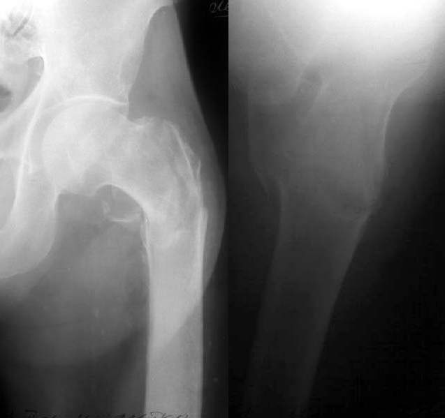
A male, 77 years old, admitted to us after a fall with the fracture (see attached). Features of the case - the patient is a pulmonary surgeon, professor and a former head of the largest pulmonary center here, still works as a scientific consultant. Hb - 77 g/l, Hct - 22%, and he doesn't allow any blood transfusion neither now nor later (). He is mentally alert but quickly gets tired. He is not obese. Very active smoker - 3 packs daily.
What would you suggest as an optimal treatment for him? We think about external fixation as a quick, easy and least invasive procedure.
|
THX in advance.
Best regards,
Alexander N. Chelnokov
Ural Scientific Institute of Traumatology and Orthopaedics
str.Bankovsky, 7. Ekaterinburg 620014 Russia
Date: Tue, 14 Aug 2001 17:57:44 -0500
From: Adam Starr
Hi Alex.
On these films, it looks like a comminuted fracture that involves the whole trochanteric region - greater and lesser trochs busted, and maybe even some extension below the lesser trochanter, into the subtroch region.
I would treat it with an intramedullary device like a Richards' IMHS or a Howmedica Long Gamma Nail. I would put the patient supine on a fracture table with the affected leg in traction, the affected hip adducted (a lot) so the trochanteric entry is easier.
Traction will probably align the fracture pretty well. Sometimes the distal fragment will sag posteriorly - be sure to check it on a lateral image view before you pass the guide wire and ream. You can correct this by lifting it up with your hand or a bone hook. You might even be able to rig a little bolster attached to the bed that would keep it held in place. That way you wouldn't have to hold it the whole time.
The proximal fragment sometimes flexes up. You can correct this by pushing down on the proximal fragment with a ball spike or a Cobb elevator. You'll need to perform these reduction maneuvers each time you pass the reamer to make sure it passes down the middle of the canal.
The nice thing about the IMHS and the long gamma nail is that they can be placed through small incisions, quickly, and you usually don't lose a lot of blood. Maybe not as minimally invasive as your planned ex-fix, but almost. And, I bet an intramedullary device would offer a better reduction and better stability. Both the IMHS and gamma nail allow immediate mobilization.
We don't treat a whole heck of a lot of geriatric patients here at Parkland, but we do see some, and we've had good results with these 2 devices.
Maybe Mike Baumgaertner or Ken Koval can give you some better advice. Or at least more learned advice. They've both written extensively on the treatment of hip fractures in this patient population.
If you do treat it with an ex-fix, be sure and post the result. I'd be curious to see what it looks like.
Good luck.
Adam Starr
Dallas, Texas
Date: Wed, 15 Aug 2001 21:33:37 -0400
From: Amir Matityahu
Dr. Chelnokov,
In my institution we have a large geriatric population. Things to consider pre and post op are the medical status of the patient as well as the nutritional status. A lymphocyte count less than 1500 and a low albumin level may be poor determinants of post op mortality and length of hospital stay (Koval KJ. Maurer SG. Su ET. Aharonoff GB. Zuckerman JD. The effects of nutritional status on outcome after hip fracture. Journal of Orthopaedic Trauma. 13(3):164-9, 1999 Mar-Apr.(Zohman GL. Lieberman JR.Perioperative aspects of hip fracture. Guidelines for intervention that will impact prevalence and outcome. American Journal of Orthopedics 24(9):666-71, 1995 Sep.)
Factors associated with poor outcome in hip fractures include poor nutrition, impaired mental ability and mobility, age, male sex, and inadequate fluid resuscitation and medical stabilization prior to surgery. Unfortunately, you can't do anything about this acutely. My major question is why is this patient so anemic? Hct of 22%? In my institution, this patient would get either a perc. dynamic hip screw or a gamma nail. Lately, we have done more short and long gamma nails as our operative time is short (20-25 minutes). Theoretically, the IM nail has better biomechanical advantage due to shorter lever arm. However, we have not had breakage of the DHS using the Howmedica Omega hip screw at the screw plate interface. The patient would get perioperative antibiotics and this would be done on a fracture table. Reduction is achieved pre-op under image and if need be, we place a well padded crutch under the hip for posterior sag.
I can't comment on blood loss. It seems to be minimal. However, there is reaming involved with insertion of the nail. We adhere to the tip-apex distance as per Baumgaertner (JBJS 77A, 1995) and allow patients to be weight bearing as tolerated post op. These patients are all placed on Iron and Colace, nutritional supplements, and multivitamins in hopes this will help their nutritional status (no data). For this patient, I would work up the anemia and give Epogen pre and post op. Use hypotensive anesthesia if he can tolerate it.
We have no experience with an ex-fix with these fractures.
Date: Thu, 16 Aug 2001 11:41:23 +0600
From: Alexander Chelnokov
Hello All,
THX for all your suggestions.
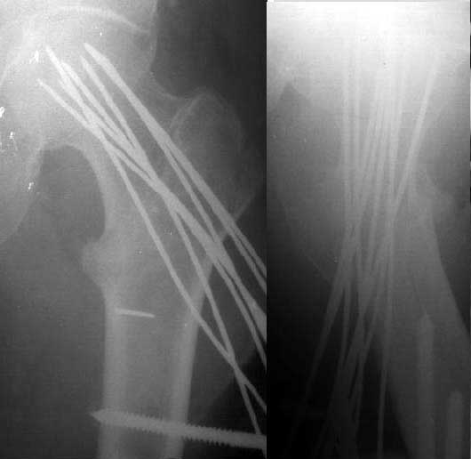
Most of the mentioned is unavalable in our institution :(. We still have not done yet any case with Ender-like nails so didn't try it for the first time in this patient. So external fixation was performed (see attachment).
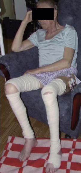
Two hours after the surgery the patient was sitting and happily smoking . Yesterday he tried to walk with crutches, but he is too weak for this, he can stand only. This morning he left for home.
 |
 |
Best regards,
Alexander N. Chelnokov
Ural Scientific Institute of Traumatology and Orthopaedics
str.Bankovsky, 7. Ekaterinburg 620014 Russia
Date: Thu, 16 Aug 2001 10:57:38 -0500
From: Adam Starr
Hi Alex.
THere's more than one way to skin a cat, I guess. THe reduction looks fine on those films. It's too bad you couldn't get a picture of him smoking - that would've been great.
What sort of weight bearing guidelines did you give him? Do you try to limit his weight bearing, or is he allowed to do whatever he wants?
Thanks for posting the post-op images. I've never seen anything like that before. It looks pretty nice, actually. Nice case. Y'all are the Ilizarov kings.
Adam
Dallas
Date: Sat, 18 Aug 2001 10:39:42 +0600
From: Alexander Chelnokov
Hello Adam,
AS> looks fine on those films. It's too bad you couldn't get a picture of him smoking - that would've been great.
:-) As a lecturer he would avoid such publicity.
AS> What sort of weight bearing guidelines did you give him? Do you try to limit his weight bearing
Weight bearing at painless level is recommended.
AS> Thanks for posting the post-op images. I've never seen anything AS> like that before.
It is difficult to show something unusual to so advanced and educated ortho people :-). Actually the approach seems to be unnesessary if one has available PFN, gamma nail and DHS. But maybe it could be useful for some indications even in this environment.
AS> It looks pretty nice, actually. Nice case. Y'all are the Ilizarov kings.
Really it doesn't require high skills. If one is able to perform proper reduction the rest is easy. Your placement of long screws from acetabular roof to pubic rami looks much more impressive.
Best regards,
Alexander N. Chelnokov
Ural Scientific Institute of Traumatology and Orthopaedics
str.Bankovsky, 7. Ekaterinburg 620014 Russia
Date: Sat, 18 Aug 2001 11:07:54 +0600
From: Alexander Chelnokov ase> To what are these numerous wires attached ? Would you please show
the whole frame you used.
The frame is pretty simple, just a plate with holes and few posts. I
haven't prepared a detailed view of the device in this patient so
found the attached example from another case.
Best regards,
Alexander N. Chelnokov
Date: Sat, 18 Aug 2001 11:35:56 +0600
From: Alexander Chelnokov
Hello Bill,
BB> You should write up a series of these ex-fixes. As Adam
says, I don't think there is anything like it in the literature over here.
Maybe.
BB> What is the complication rate re infection
Not outstanding, usually superficial, mostly due to skin cut out
by wires.
BB> and fixation failure?
Never met.
BB> How long do you have to keep the fixator on?
Depends on fracture pattern and quality of reduction. From 4 weeks to
5-6 months. Most cases about 8 weeks.
BB> What special postop precautions??
Can't remember something special. Painless weight-bearing, watch for
tissue conditions, keep knee ROM.
Best regards,
Alexander N. Chelnokov
Date: Sat, 18 Aug 2001 21:07:55 +0600
From: Alexander Chelnokov
Hello,
DTM> Are these wires are held strong enough to prevent dispacement?
Hardly ever eight 2 mm wires give stability for long distance run
but it is enough for common postoperative management with limited
weight-bearing. Anyway the approach never leads to something like DHS
cut out. Also less number of 3 mm Steinman pins or 6 mm half pins can
be used if the bone quality allows.
DTM> And what is about pin tract infection which may involve
fracture
Never met such a complication. Infection is usually superficial and is
developed after some weeks so if oral ciprofloxacin is not enough to
control it within 2 days, the involved wire can be removed.
DTM> as well as hip joint.?
I remember two such cases about 10 years ago in rather young patients
with neck, not throchanteric, fractures- both were featured with
transarticular wire placement and very long fixation (more than 6
months).
Best regards,
Alexander N. Chelnokov

Ural Scientific Institute of Traumatology and Orthopaedics
str.Bankovsky, 7. Ekaterinburg 620014 Russia
Ural Scientific Institute of Traumatology and Orthopaedics
str.Bankovsky, 7. Ekaterinburg 620014 Russia
Ural Scientific Institute of Traumatology and Orthopaedics
str.Bankovsky, 7. Ekaterinburg 620014 Russia