
Date: Wed, 10 Oct 2001 14:20:12 +0600
Subject: Femoral head fx
Hello all,
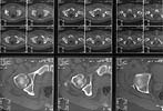
A male 25 y.o., admitted to us at 10th day after MVA. The hip luxation was reduced at the initial hospital acutely. The question is about his femoral head fracture (AP view and CT attached). How to manage this lesion? Leave "as is"? ORIF? The fragment removal?
 |
 |
THX for your opinions.
Best regards,
Alexander N. Chelnokov
Ural Scientific Institute of Traumatology and Orthopaedics
str.Bankovsky, 7. Ekaterinburg 620014 Russia
Date: Wed, 10 Oct 2001 07:33:27 -0700
From: Chip Routt
Hello Alex-
As you may know, this is insufficient imaging provided for us to make treatment recommendations. No one would treat a patient with this type of unusual injury based on a single AP hip plain radiograph and a single axial image low through the joint.
Perhaps Judet obliques, and pelvic/acetabular zone CT scan images would reveal complete information to make a good plan.
But, maybe there are some "seers" on the list.
Chip
Date: Wed, 10 Oct 2001 09:52:18 -0500
From: Steven Rabin
No, I also held off giving an opinion because I think we need more information.
Date: Wed, 10 Oct 2001 12:08:38 -0600
From: Thomas A. DeCoster
I have left small fragments from the femoral head and patients seemed to do OK. This one appears to be relatively inferior and is not causing appreciable widening or malreduction of the hip joint.
Fixing them is, as you know, difficult. The surgical approach, visualizing both the fragment and the defect site, obtaining reduction, obtaining and maintaining fixation, devascularizing the fragment or the femoral head,... are all problems.
If you go posterior and redislocate the hip the fragment tends to stay in front. If you go anterior you must further disrupt the blood supply to the femoral head. Absorbable pins recessed beneath the articular surface can provide sufficient stability in some cases.
TDeCoster
Date: Wed, 10 Oct 2001 13:36:38 -0700 (PDT)
From: mahmoud al-salhi
Hello All,
My feeling is to leave this fragment, and it seems still attached and separated 10 days after the injury. But my question is, can arthroscopy help in these cases?? Keeping in mind that this fracture is in a non-weight bearing area.
Date: Wed, 10 Oct 2001 18:31:48 -0700
From: Chip Routt
It would be nice to see the complete injury films and images.
I'm curious if anyone on the list has ever accurately reduced and fixed an anteromedial displaced femoral head fracture using a posterior exposure.
Is there really evidence/information that an anterior surgical exposure of the hip in such a patient "further disrupts" the femoral head blood supply?
Also, has anyone ever "performed" a hip arthroscopy soon after such an injury? How did that go? Please comment on inflow.
Maybe more images?
Chip
Date: Thu, 11 Oct 2001 06:50:19 -0500
From: Adam Starr
I've never reduced and fixed one using a posterior approach. I've always relied on a Smith Peterson approach. And on most occasions, I've been happy to find that the anterior, low fragment still has a little strip of periosteum attached to it.
I've stuck my finger around the bottom of the neck to reach the front on a couple cases where we had tiny anterior fragments that we decided to ignore. You can move them a bit, but I don't think I could say I "reduced" them.
Adam Starr
Dallas
Date: Thu, 11 Oct 2001 17:13:33 +0000
From: b.meinhard
Not Proud, but I have reduced a devitalized piece from the rear, and this necessitated an open redislocation and capture by hardware from back to front.
bpm
Date: Thu, 11 Oct 2001 17:52:39 -0400
From: Charles Blitzer
I have had the opportunity to 'scope a couple of hips post reduction to remove loose bodies. I used gravity inflow and did not have any problems. No serious extravasation.
Charles Blitzer
Date: Thu, 11 Oct 2001 15:43:18 -0700
From: Chip Routt
could you share those pre- and postop CTs?
Chip
Date: Fri, 12 Oct 2001 00:22:08 +0600
From: Alexander Chelnokov
Hello Chip,
CR> unusual injury based on a single AP hip plain radiograph and a single axial image low through the joint.
I picked most clear views of the injury to my mind. At the moment available are one Judet view and, more CT scans and AP view of the pelvis where i cut the image from - attached to this and the next message. Quality of some initial films with luxation and after reduction is below than acceptable level.
CR> reveal complete information to make a good plan.
I hope so. THX!
 |
 |
 |
Best regards,
Alexander N. Chelnokov
Ural Scientific Institute of Traumatology and Orthopaedics
str.Bankovsky, 7. Ekaterinburg 620014 Russia