

Date: Fri, 22 Nov 2002 18:52:15 +0500
Subject: Femoral neck fracture
Hello All,
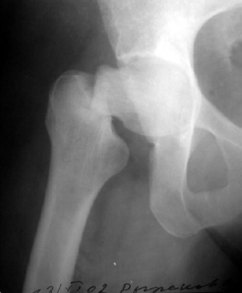
A female 40 y.o. transferred to us from elsewhere. Two weeks ago she stumbled at home. She reports conservative treatment of the hip arthritis, no previous x-rays presented. She has been marked pain in the hip recent months. Xrays demonstrate no sharp end. Isn't it a fatigue fracture?
Add also her obesity. She remembers no hip ROM limitation prior the accident.
|
|
|
Our opinions diverge from cancellows screw fixation to THR, with medializing/lateralizing osteotomy in the middle.
Pls help with a rationale for the choice.
Best regards,
Alexander N. Chelnokov
Ural Scientific Institute of Traumatology and Orthopaedics
str.Bankovsky, 7. Ekaterinburg 620014 Russia
Date: Fri, 22 Nov 2002 09:29:04 -0500
From: James Carr
To my eye, it does have a sclerotic edge to it, but it may just be projection . Does she have diabetes or other metabolic - eg renal problems?
Jim Carr
Date: Fri, 22 Nov 2002 20:36:07 +0530
From: Dr Mangal Parihar
Alex,
Rather than a fatigue fracture, I would look for a pathological fracture and rule out either metastases or metabolic disease.
If she reports no limitation of hip movements before the injury, then I would not think of arthritis in the hip, and it would be even more important to rule out the differentials of mets or metabolic disease
If it is mets, then the decision may change, but otherwise, internal fixation with multiple screws would be the way to go, One could either do a closed reduction (which should not be a problem in a basal fracture like this one, or an open reduction if one cannot achieve a closed reduction.
Certainly, a thr in this patient would be overkill - reasons - young patient & overweight both of which will lead to earlier failur thru wear...
regards
Date: Sat, 23 Nov 2002 18:26:49 -0500
From: Fred Barrick
It looks like there is an early fatigue (stress) fracture of the opposite hip as well. I would fix both hips with screws, then get a bone mineral density and other endocrinologic work-up
Fred Barrick MD
Inova Regional Trauma Center
Falls Church, VA
Date: Sun, 24 Nov 2002 21:42:55 +0530
From: DR T I GEORGE
Dear Alex,
As suggested by many members, there seem to be something more than just a one sided neck of femur fracture.
Will be happy to know her biochemical work up.
Do you have an x-ray skull lateral view??
DR T I GEORGE
Date: Mon, 25 Nov 2002 18:14:08 -0500
From: David Goetz
Looks like a displaced stress fx to me. Have been sent many over the years and and seen [and contributed] to a 100% failure rate when treated with just cannulated screws and an indirect reduction. Screws will work in the acute situation, but not chronic. The reduction of a delayed fx can never be truly anatomic. I would suggest an osteotomy along with a blade plate or compression screw or at least a compression screw in anatomic position or slight valgus with normally aligned medial cortex [hard to do].
David R. Goetz MD
Medical Director, Orthopaedic Trauma, Greenville SC
Date: Tue, 3 Dec 2002 00:54:23 +0500
From: Alexander Chelnokov
Hello All,
The patient fell Nov 5. And some months prior the episode she marked pain in the hip. And "something was wrong" with the hip for about 2 years. As she reports, there was no hip ROM decrease.
Today her film was received dated Nov 1, four days before the accident - attached.
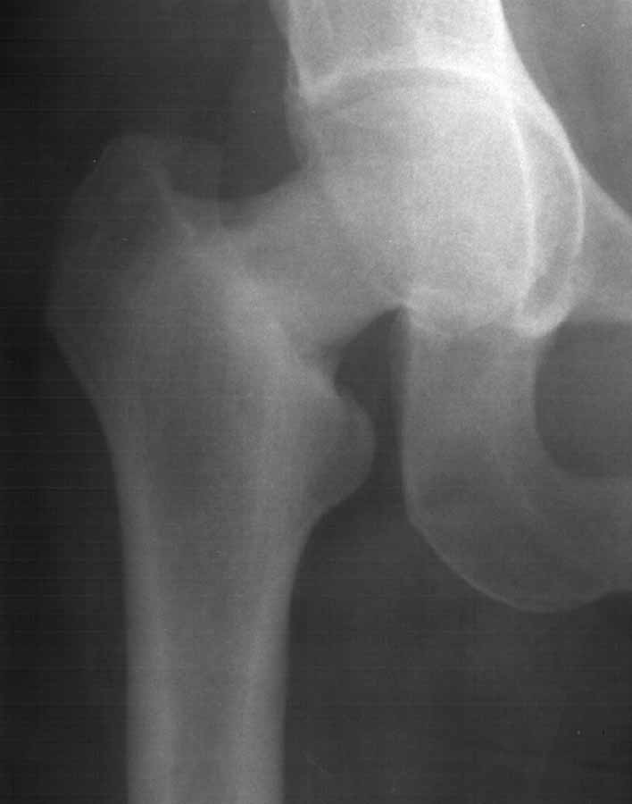 |
Maybe it is still fatigue fracture?
Best regards,
Alexander N. Chelnokov
Ural Scientific Institute of Traumatology and Orthopaedics
str.Bankovsky, 7. Ekaterinburg 620014 Russia
Date: Mon, 2 Dec 2002 17:24:25 -0600
From: Sciadini, Marcus
CT scan available?
MFS
Date: Sat, 7 Dec 2002 17:34:55 +0500
From: Alexander Chelnokov
Hello All,
sak> Have you ruled out all forms of metastatic bone disease (more so CA breasts)? The head and neck look pretty sclerosed to me.
She was operated this Thursday. Closed reduction failed. When the site was exposed it appeared very sclerotic, almost not bleeding. Well, we reduced fragments and transfixed with temporary wires. After this a cancellous screw was inserted to the upper pole. While a seating chisel was being inserted the femur cracked despite it was predrilled. :-( There was no implant suitable for the new pattern. So we took a DCP-like plate, overdrilled two upper holes and modelled it. The neck was fixed with two more 6.5 mm screws. Image attached.
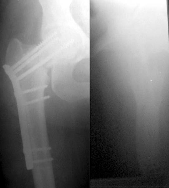 |
The bone really was very sclerotic. What is a possible cause of this? It doesn't look like pure Padgets or a metastatic case.
Best regards,
Alexander N. Chelnokov
Ural Scientific Institute of Traumatology and Orthopaedics
str.Bankovsky, 7. Ekaterinburg 620014 Russia
Date: Sat, 7 Dec 2002 9:37 AM EST
From: Bill Burman
Alex
There is a discussion of osteopetrosis (possibly a tarda variant in this case) at the emedicine.com web site which includes a good differential diagnosis of osteosclerotic lesions. i.e.
Date: Mon, 9 Dec 2002 11:37:19 -0700
From: Thomas A. DeCoster
Alex,
I totally agree with Bill Burman's suggestion that the etiology of this pathologic process should be pursued aggressively. The list of possible diagnoses he provided and the URL link are a good start on a diagnostic workup. With a pathological fracture primary emphasis should be on the pathological condition and secondary empahsis on the fracture and fixation type. Your fixation will probably hold up long enough to make a pathological diagnosis. Treatment of the pathological condition is then based on the diagnosis and ultimately, probably more important than the fracture treatment itself. Fracture treatment may also be more a function of the pathological entity than the fracture pattern.
Osteopetrosis tarda seems to me to be one reasonable possible diagnosis to pursue.
TD