Date: Thu, 20 Mar 2003 23:21:15 -0600
Subject: 5 months S/P Pelvic Injury
Dear All,
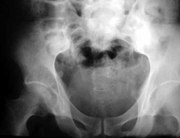
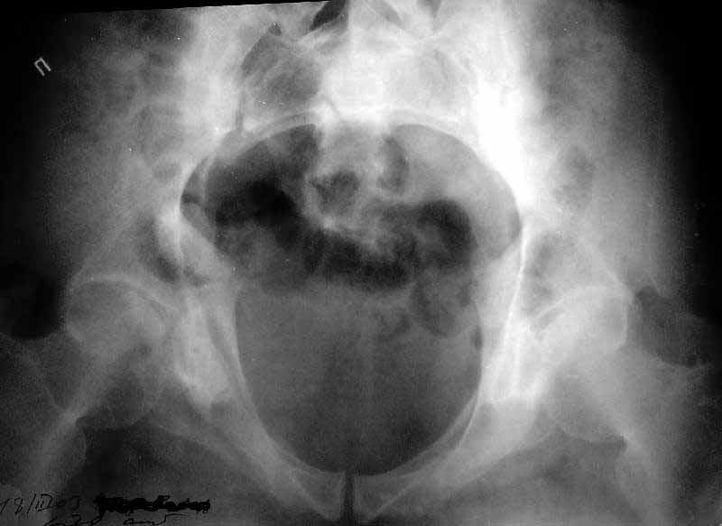
A male 64 y.o., physical worker, Oct 29, 2002 sustained work related anterior blunt impact by a massive tube. He was treated somewhere else by 2 months bed rest, then 2 months with crutches. Now he walks with cane and experiences pain in legs and limp. Often nocturnal pain, daily uses analgetics. X-rays show unreduced posterior lesion. What would you do with the patient?
|
|
THX in advance.
Best regards,
Alexander N. Chelnokov
Ural Scientific Institute of Traumatology and Orthopaedics
str.Bankovsky, 7. Ekaterinburg 620014 Russia
From: Bill Burman
Alex,
where is the pain and tenderness? does it radiate to both legs or is it just in both legs?, neurologic exam ?, sciatic tension signs? Patrick/Fabere test?, Gaenslen's sign? leg length discrepancy? any outlet view ? posterior pelvic-sacral CT cuts?
Date: Fri, 21 Mar 2003 07:47:02 -0800
From: Chip Routt
What would you do with the patient?
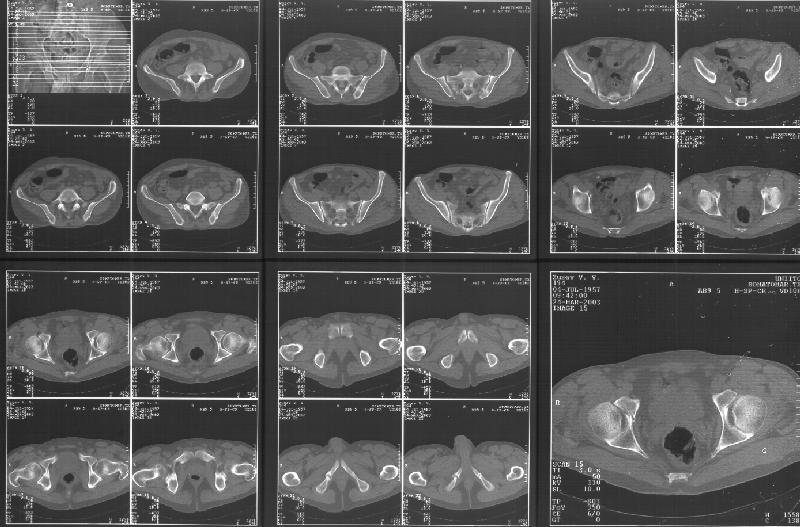
A pelvic CT scan.
Chip
M.L. Chip Routt, Jr.,M.D.
Professor-Orthopedic Surgery
Harborview Medical Center
Seattle, WA 98104-2499
Hello All, especially Bill and Chip,
CR> A pelvic CT scan.
Attached. I may upload the one with larger resolution.
 |
Any suggestions?
Best regards,
Alexander N. Chelnokov
Ural Scientific Institute of Traumatology and Orthopaedics
str.Bankovsky, 7. Ekaterinburg 620014 Russia
Date: Wed, 26 Mar 2003 06:48:22 -0800
From: Chip Routt
Hello Alex-
Alternating single leg stance AP pelvic films ("flamingo views") may be helpful to identify hemipelvic instability.
M.L. Chip Routt, Jr.,M.D.
Professor-Orthopedic Surgery
Harborview Medical Center
Seattle, WA 98104-2499
Date: Wed, 26 Mar 2003 18:59:22 -0700
From: Terry Finlayson
Based on the CT images you provided, he appears to have a nonunion of the the right ischium, which would explain his symptoms. General principles of treatment of hypertrophic (which this appears to be) nonunions suggest rigid fixation, but I'm not sure what the best approach would be. Could get a lag screw across the fracture through a Kocher-Langenbeck approach or plate osteosynthesis through direct ischial approach, but perhaps Chip, Adam or someone with more pelvic fracture experience could enlighten us all.
Terry Finlayson, MD
Alpine Orthopaedic Specialists
Logan, UT
Date: Wed, 26 Mar 2003 19:51:50 -0800
From: Chip Routt
I didn't see it on the images...maybe I need glasses!!
I'll try to enlarge the images to understand.
M.L. Chip Routt, Jr.,M.D.
Professor-Orthopedic Surgery
Harborview Medical Center
Seattle, WA 98104-2499
Date: Thu, 27 Mar 2003 12:31:50 -0600
From: Adam J. Starr, M.D.
I didn't see it either - can anybody re-send me the CT images?
Presuming the patient DID have a nonunion of an ischial fracture, I'm not sure what the right course of treatment would be. You'd have to make yourself pretty darn sure that the symptoms were actually coming from the nonunion...and sometimes pain around the hip/butt is hard to pinpoint.
If you decided that the pain and symptoms WERE due to a nonunion, then you'd have to talk with the patient about whether the treatment to fix the nonunion would be worth it. Open reduction, plating and possible bone grafting of the ischium (presumably via a Kocher) is a pretty big operation. The guy might decide the treatment is worse than the disease.
The option of percutaneous stabilization exists at a few centers, but I don't know that anybody has a big series of pelvic nonunions treated percutaneously so that we can say "this method works". There are a couple case reports in the literature.
I've done a number of sup ramus nonunions, and they seem to work okay. I've treated one transverse acetabular fx nonunion percutaneously, and it worked - pain went away and the fx healed. But, that's just a handful of anecdotes.
Adam Starr
Dallas, Texas
Date: Thu, 27 Mar 2003 15:37 EST
From: Bill Burman
I think the cuts in question (albeit small) are on the right of the bottom row of the CT scan
Date: Thu, 27 Mar 2003 17:38:51 -0600
From: Adam J. Starr, M.D.
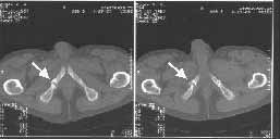
I think this is just a pelvic ring disruption that has gone on to a non-union. The arrow is pointing to the inferior ramus non-union, and the sup ramus is a high "root" fracture, right near the pecten. The sup ramus fracture enters the tab - you could call it an acetabular fracture - but down here in Dallas we treat these acutely as stable pelvic ring injuries. The fracture in the anterior portion of the acetabulum is so low it doesn't seem to cause much trouble.
His R sacroiliac joint is opened anteriorly a little bit - the "unreduced posterior lesion" Alex mentioned in his first post. My earlier post about plating, bone grafting, etc., is all wrong.
What you have is a pelvic fracture that hasn't healed after 4 months or so, with a minimal deformity.
I think what I would offer the guy is a percutaneous iliosacral screw to stabilize and improve the SI joint alignment, coupled with a perc anterior column screw to stabilize the high ramus fx. These screws aren't easy to place, and have a host of potential problems and complications. But, I think they would work.
The open treatment options carry more morbidity, in my opinion, but they could achieve the same result. It's possible to do an ilioinguinal approach and stabilize both fractures. Bone grafting of each site (you would probably aim for a fusion of the SI joint if you chose the open route) and plate fixation would likely lead to union.
The perc screw method would also likely lead to union, but with less surgical dissection. The problem with the perc method is that it requires an expert fluoroscopy technician, and a surgeon who understands the anatomy, as seen on fluoro. It's hard to do.
Adam Starr
Dallas, Texas
Date: Fri, 28 Mar 2003 17:12:42 +0500
From: Alexander Chelnokov
Here is a link to "full-size" CT image (scanned with 300 dpi resolution to 2776x1832 pixels). Thanks for advice.
Best regards,
Alexander N. Chelnokov
Ural Scientific Institute of Traumatology and Orthopaedics
str.Bankovsky, 7. Ekaterinburg 620014 Russia
Date: Fri, 28 Mar 2003 09:05:02 -0600
From: Kyle Dickson, MD
Cannot define the deformity completely with the limited views available. The problems with pelvic pain after injury is try to define where the pain is coming from which is very difficult to do. Selective injections with lidocaine under flouro can help (si joint, rami fx, etc.). the problem with percutaneous fixation is you go against a personal orthopaedic principle, "Don't fix a malunion" this will only get you into trouble down the road.
kyle