
Date: Tue, 6 Mar 2001 12:06:26 +0500
Subject: Femoral Interlocking Nail
From: Alexander Chelnokov
Hello All
A first case of the closed subject has just been performed in our unit (guess who :-). The nail was a rectangular flat titanium plate 12 mm wide and 5 mm thickness (from a provision set for osteosynthesis by individually modelled nails according to a rather popular in Russia methodic of Zverev-Klyuchevky). It was only bent in sagittal plane and its ends were rounded. Cortical 4,5 mm titanium screws were used for locking performed as free-hand at both levels.
The case was selected because the whole isthus zone was comminuted and no reaming was required - no appropriate equipment is available. What was reached can be seen on attached x-rays.
|
As nobody here has any personal experience in the approach (actually there was no case of 1)closed and 2)interlocked femoral nailing for thousands kilometers around) a strong skepticism was a reaction on the surgery, particularly from our vice-director.
I would like to hear your opinion about perspectives of union in the case. Is the quality of reduction acceptable for this kind of injury and operative technique? Or we should open the fracture site and perfrom additional reduction?
The patient has just left the clinic with painless knee motions and toe-touch wight-bearing on 7th postoperative day.
THX in advance.
Best regards,
Alexander N. Chelnokov,
Ural Scientific Institute of Traumatology and Orthopaedics
Ekaterinburg 620014 Russia
Date: Tue, 6 Mar 2001 07:41:04 -0000
From: chris wilson
The entry point of the nail is a little medial, so the most proximal fragment is in varus. That said, it's a pretty good position for such a difficult fracture, especially done closed. In our institution, the position would be deemed most acceptable, and we would be prepared to wait 3 months-if there was no callus at all then, we would bone graft it.
Chris Wilson
Knee and Trauma Surgeon
University Hospital
Cardiff, UK
Date: Tue, 6 Mar 2001 04:57:45 -0800
From: bruce meinhard
Alex,
Alignment good, but one really needs to see the joint above and joint below to be sure. High probability of healing without additional surgeries especially if the fracture was not surgically opened and stripped of blood supply. This can be approached from supine position or with the patient in lateral decubitus position.
BPM
Date: Tue, 6 Mar 2001 17:31:08 +0500
From: Alexander Chelnokovts.ru>
Hello chris,
cw> The entry point of the nail is a little medial, so the most proximal fragment is in varus.
I intentionally inserted the nail medially to prevent varus so it seems to me it must be valgus maybe?
cw> That said, it's a pretty good position for such a difficult
THX for your support!
Best regards,
Alexander N. Chelnokov,
Ural Scientific Institute of Traumatology and Orthopaedics
Ekaterinburg 620014 Russia
Date: Tue, 06 Mar 2001 06:53:58 -0600
From: Adam Starr Alex,
Good for you! Nice job.
My prediction is that you'll see abundant callus within a month or so, and that it will unite uneventfully. Reaming would've helped this occur faster, but I bet it goes ahead and heals anyway.
Be sure and give us some follow up.
Maybe I can convince one of the local intramedullary nail companies to send y'all a set, along with some reamers. Do you have a fluoroscopic C-arm?
Adam Starr
Dallas, Texas
Date: Wed, 7 Mar 2001 00:55:15 +0500
From: Alexander Chelnokov
Hello Adam,
AS> My prediction is that you'll see abundant callus within a month or so, and that it will unite uneventfully.
THX for the prognosis!
AS> Reaming would've helped this occur faster, but I bet it goes
I mentioned reaming to explain only why i selected the comminuted case for the first time. In a simple fracture i supposed a high risk of shaft splitting by the wide nail, but the one had nothing to split more so reaming was not needed at all.
AS> Be sure and give us some follow up.
Sure.
AS> Maybe I can convince one of the local intramedullary nail companies to send y'all a set, along with some reamers.
Sounds great.
AS> Do you have a fluoroscopic C-arm?
You strongly overestimate my skills if suppose that i am able to perform closed insertion of such a nail and its free-hand locking without fluoroscopic control :-)
Alexander N. Chelnokov,
Ural Scientific Institute of Traumatology and Orthopaedics
Ekaterinburg 620014 Russia
Date: Tue, 06 Mar 2001 15:49:49 -0500
From: Michael S. Sirkin, M.D.
Alignment looks good from what can be seen. With these comminuted fractures you need to sure proper length was obtained. I do this in the OR with fluoroscopy but post op scanogram either with plain X-ray or CT can be done.
Date: Tue, 06 Mar 2001 20:31:45 -0600
From: Steven Rabin
i also think this will heal fine. i agree with previous comments, but would also stress the need to check rotational alignment as well. For that, good x-rays of the hip and knee, not shown here, would be very helpful. I like to get a perfect lateral of the knee with the c-arm with the condyles overlapping and the patella straight anterior, and it should match a perfect lateral of the hip. Then a perfect AP view of the knee with the patella perfectly centered should match a perfect AP view of the hip judged by looking at the relationship/appearance of the trochanters....
good luck. i think you will have a good result.
Date: Sat, 10 Mar 2001 08:32:26 -0800
From: Carlo Bellabarba > I would like to hear your opinion about perspectives of union in the
case. Is the quality of reduction acceptable for this kind of injury and
operative technique?
Looks great, and should heal with acceptable alignment. In the relatively
low likelihood that it does not heal, having established appropriate
alignment, and presumably length and rotation, you have simplified any
potential future procedure.
> Or we should open the fracture site and perform additional reduction?
absolutely not.
simply put, you were right and your vice-director is wrong.
Carlo Bellabarba, MD
Date: Fri, 16 Mar 2001 13:49:59 -0800
From: Thomas A. DeCoster
I would re-iterate what has been said. That cases with an Xray as shown
typically heal with abundant callus and good function in a few months;
although not always and you can't be sure with one xray and the xray doesn't
give information on the soft tissue injury or dissection.
I find it fascinating that the "miracle" of intramedullary fixation is not
"available" (reamers, fluroscopy, etc) or "accepted" (vice-director,...) in
places that clearly have a generally high level of ability to care for
patients. I suppose it's similar to the "west's" reluctance to implement the
"miracle" of distraction osteogenesis. Perhaps it has nothing to do with
geography or politics.
Tom DeCoster
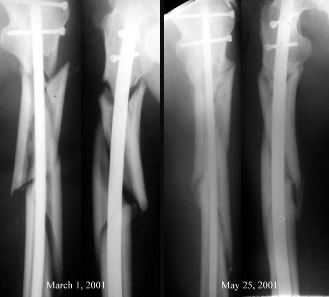
Date: Fri, 25 May 2001 20:18:10 +0600
From: Alexander Chelnokov
Hello All,
Recently i asked your opinion about a case of interlocked femoral
nailing case which was the first one in our institution. Postoperative
xrays were ugly so our vice-director insisted that open reduction was
necessary.
Comments from list members helped to avoid this procedure. Today is
about 3 months after the surgery and i am glad to inform that
all who guessed good outcome for the case won :-)
See attached images.
Best regards,
Alexander N. Chelnokov,
Spine Trauma and Reconstruction
Orthopaedic Trauma
University of Washington/Harborview Medical Center
Seattle, WA

Ural Scientific Institute of Traumatology and Orthopaedics
Ekaterinburg 620014 Russia