Date: Mon, 23 Jun 2003 13:22:40 -0500
Subject: Femoral nonunion and ?
From: Alexander Chelnokov
Dear All,
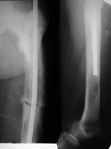
A male 76 years old sustained femoral shaft fracture after minimal
injury. Open reduction and intramedullary nailing was performed
somewhere else. He was refferred to us with pain in the femur and sense
of movement at the site, especially with rotation. Xrays revealed except
nonunion some bone structue changes. What is this? Isn't it Paget's
disease?
We plan to perform exchange closed locked nailing. How the bone status
should influence the plan? Either use a small diameter nail without
reaming at all or it would be no problem to ream to 11-12 mm?
THX in advance.
Best regards,
Alexander N. Chelnokov
Ural Scientific Institute of Traumatology and Orthopaedics
str.Bankovsky, 7. Ekaterinburg 620014 Russia
Reply at: Orthopaedic
Trauma Association forum
Date: Mon, 23 Jun 2003 14:46:06 -0400
From: James Carr
I believe its Pagets. The bone looks larger than normal, along with
coxa vara, which is characteristic. Check his alkaline phophatase. In
rare instances, you can see aggressive lytic disease (unlikely here),
which requires medical Rx to quiet down. Get out your reamers, and put
a big nail in him- the biggest his canal will take. Make it longer, and
lock if you don't believe the fixation is snug.
James B. Carr, MD
Palmetto Health Orthopedics
Date: Mon, 23 Jun 2003 14:46:01 -0500
From: Frederic B. Wilson, M.D.
Alex,
From the appearance of the x-rays and the description of a fracture after
minimal trauma, I would be very concerned about a neoplastic process. Odds
would favor something metastatic and appearance might suggest a combined
blastic-lytic lesion. I am particularly concerned about the lytic appearance
distal to the end of the rod. I would work up the patient for both neoplastic
and metabolic process before proceeding. After diagnosis treatment options
can be considered. Dying to know what this is.
- Frederic B. Wilson, M.D.
- Assistant Professor
- Trauma and Adult Reconstruction
- Department of Orthopaedic Surgery
- Tulane University School of Medicine
- New Orleans, LA, 70112
Date: Mon, 23 Jun 2003 22:15:52 +0100
From: Chris Wilson
The femur is Pagetic.
Check serum Calcium,Urea and Electrolytes and Ak Phos.Pagets may be in
active "lytic" phase.
Aim to use a strong reamed locked nail,as the femur is bowed and union
may be slow.
Be prepared for greater than usual blood loss.
Regards
- Chris Wilson
- University Hospital
- Cardiff
- UK
Date: Tue, 24 Jun 2003 16:04:54 +0530
From: DR T I GEORGE
Dear Dr Alex,
I think I will add my few thoughts.
In case this case is Paget's, be prepared for a tough time during reaming.
Bone tends to be hard and reamers may be found wanting in efficiency.
Hence keep an option for plating in case re-nailing turns out to be a tough
proposition during surgery.
Best of luck and do keep us posted on what you do.
DR T I GEORGE.
Head of Orthopaedics Unit III
Little Flower Hospital,
Angamaly,Kerala State,
India.
Date: Wed, 25 Jun 2003 11:28:38 +0600
From: Alexander Chelnokov
Hello All,
THX to all colleagues for their opinions. Chest xrays are clear. Our
radiologists explored films and swear the bone changes are Paget's.
Finally we proceeded with a minimal plan - the existed nail was pulled
out (quite easily). It turned out that a largest available nail was only
11 mm 40 cm, so i inserted it and locked dynamically. The procedure was
quick. As immediate effect the patient marked lack of clicking which
disturbing him before. We plan to discharge him ASAP with
recommendations of biphosphonates etc.
Best regards,
Alexander N. Chelnokov
Ural Scientific Institute of Traumatology and Orthopaedics
str.Bankovsky, 7. Ekaterinburg 620014 Russia
Date: Thu, 26 Jun 2003 14:50:26 -0600
From: Thomas A. DeCoster
It looks like Paget's disease to me. The original nail is small, short and
unlocked. Make the second one reamed, large (14 mm), long (past the distal
lucency) and statically locked for rotational control. Send tissue for
pathology.
Tom DeCoster