
Date: Mon, 4 Aug 2003 17:55:34 +0600
Subject: Distal tibial fixation failure
Dear All,
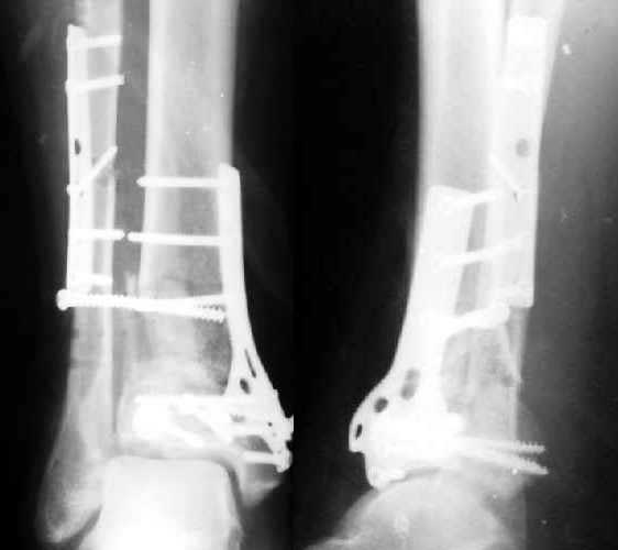
6 weeks ago a pilon fracture in a male 45 years old was ORIFed (image 1). The patient also had the opposite foot injured. Due to urological problems he then was transferred to another facility. Against our advice he walked with partial weight-bearing. Partial loss of fixation was revealed when a month later he visited our unit (image 2).
|
|
He is also in waiting list for repair of the urethra and now with the urinary fistula. What options do exist for the re-displaced ankle?
THX in advance.
Best regards,
Alexander N. Chelnokov
Ural Scientific Institute of Traumatology and Orthopaedics
str.Bankovsky, 7. Ekaterinburg 620014 Russia
Date: Mon, 4 Aug 2003 08:19:37 -0400
From: James Carr
Alex
Is he diabetic (or pre diabetic and doesn't know it??. I would go lateral through fibular incision - assuming healing, and plate from lateral. I would also pay attention to the syndesmosis, as this may require some fixation. If he is a diabetic, the issue is more cloudy. I would still probably fix it. This guy then gets a bent knee long leg cast for 6 weeks postop.
James B. Carr, MD
Palmetto Health Orthopedics
Date: Tue, 5 Aug 2003 14:39:53 +0600
From: Alexander Chelnokov
Hello James,
JC> Alex Is he diabetic (or pre diabetic and doesn't know it??).
No.
JC> I would go lateral through fibular incision - assuming healing, and plate from lateral.
Wouldn't it lead to excessive devascularization of the distal tibia?
JC> I would also pay attention to the syndesmosis, as this may require some fixation.
So i keep in mind an external fixator and closed insertion of 1-2 thin bolts to compress synesmosis...
JC> would still probably fix it. This guy then gets a bent knee long leg cast for 6 weeks postop.
Or bridging ex-fix?
Best regards,
Alexander N. Chelnokov
Ural Scientific Institute of Traumatology and Orthopaedics
str.Bankovsky, 7. Ekaterinburg 620014 Russia
Date: Mon, 4 Aug 2003 13:04:14 EDT
From: Tadabq
This is a BIG problem, that fortunately is somewhat less common now than 10 years ago. The fibula and a large piece of distal lateral tibia are now displaced laterally about 1 cm. It is difficult to assess how well reduced the tibia articular surface was and is. You might consider:
1. What is the status of the soft tissues? Range from grossly infected with tissue loss to healing OK with surgical scars. Many somewhere in between but the soft tissue slough is a common and BIG problem. You haven't mentioned much about this.
2. What is the status of the patient? Diabetic?, compliant?, smoking? etc.
3. What is the status of the fixation? Is it solid or grossly loose? This appears somewhere in between with perhaps syndesmosis instability but the other fixation (fibula, tibia articular surface, tibia meta-diaphysis ?OK)
4. What is the status of the reduction. Again, syndesmosis very wide but other (fibular length, tibia articular surface, tibia alignment, talus beneath plafond) seems OK.
5. What is your risk tolerance? Individualized to patient and surgeon. How much risk of BKA are you willing to take to try to get somewhat improved ankle function.
I believe this may be salvage mode and a good ultimate result will be a preserved foot and ankle fusion. So be careful about doing too much that might result in infection, multiple operations and BKA. If soft tissue OK, patient OK, reduction and fixation failure limited to sydesmosis etc then you could try repeat syndesmosis reduction and new fixation. Perhaps 2 screws with bicortical tibia and fibula purchase supplemented by enforced NWB (bent knee LLC vs other).
TD
Date: Tue, 5 Aug 2003 15:07:44 +0600
From: Alexander Chelnokov
Hello Tom,
TAC> 1. What is the status of the soft tissues?
No problem.
TAC> 2. What is the status of the patient? Diabetic?, compliant?, smoking? etc.
He is smoker, non-diabetic, but with cystostoma and a lot of WBC in urine. Now looks compliant, at least doesn't weight-bear the leg.
TAC> 3. What is the status of the fixation? Is it solid or grossly loose? This appears somewhere in between with perhaps syndesmosis instability
Exactly.
TAC> but the other fixation (fibula, tibia articular surface, tibia meta-diaphysis ?OK)
At least he is now without external immobilization, moves to/from bed/wheelchair without sense of instability.
TAC> 4. What is the status of the reduction. Again, syndesmosis very wide but other (fibular length, tibia articular surface, tibia alignment, talus beneath plafond) seems OK.
Yes. Looks like pure depression of the grafted metaphyseal part. Of course along with separation of the lateral aspect (plus syndesmosis and tibia).
TAC> 5. What is your risk tolerance? Individualized to patient and surgeon. How much risk of BKA are you willing to take to try to get somewhat improved
I suppose here to avoid procedures where the risk can't be so small to be neglected.
TAC> OK, reduction and fixation failure limited to sydesmosis etc then you could try repeat syndesmosis reduction and new fixation.
It seems to me the articular tilt should be restored.
TAC> Perhaps 2 screws with bicortical tibia and fibula purchase supplemented by enforced NWB (bent knee LLC vs other).
Yes, the screws can be inserted through stab wounds. My concern is what to do if closed foot distraction wouldn't provide "elevation" of the articular surface.
Best regards,
Alexander N. Chelnokov
Ural Scientific Institute of Traumatology and Orthopaedics
str.Bankovsky, 7. Ekaterinburg 620014 Russia
Date: Tue, 5 Aug 2003 22:23:55 EDT
From: Tadabq
Alex,
Regarding the case of loss of reduction after plating distal tibia fracture
It sounds like we are of similar opinion on this case. I didn't notice the collapse of the metaphyseal bone graft and talar tilt but upon re-review of the radiograph I see what you mean. I haven't had ANY luck successfully repairing this situation. Half the time it still doesn't look right on the post op x-rays (?perhaps inadequate surgeon-me). Half the time it collapses later and the other half (sic) it doesn't matter because they develop some other greater problem like totally degenerative joint.
After looking at these fragments in acutely operatively treated distal tibia fractures and seeing no soft tissue attachment (all the depressed articular pieces are clearly without soft tissue attachment and many of the cortical pieces are quite alone) I wonder if a big part of the problem isn't avascularity of the fragments. When I have gone back in late, the pieces often look like unincorporated bone graft, just sitting there. Even if I restore some semblance of a normal plafond the fragments seem to behave like femoral head grafts I once used to supplement deficient acetabulae for total hips- it looks OK for a while but eventually melts away.
I'm not saying it's impossible to restore the joint and obtain a good result at this point, but I've never been successful once it gets to this stage.
There is no situation so bad that it can't be made worse by a well-meaning orthopedist.
TD
Date: Tue, 5 Aug 2003 07:37:04 EDT
From: Aobonedoc
cast him and wait. Deal with the residual at a later date. He had his chance.
Sincerely and respectively,
M. Bryan Neal, MD
Arlington Orthopedics and Hand Surgery Specialists, Ltd.
Arlington Heights, Illinois 60005