Hello Adam,
Didn't you try to explore the base of the refractures? I've never met refractures in pediatric/adolescent femurs but I deal mostly with adults so children were occasional. I asked our ped guys and they don't remeber such cases also. The only case i met was in a patient with osteogenesis imperfecta. Maybe the fixation was too stable and re-fractures were due to primary healing which is occured early but is not too strong?
Best regards,
Alexander N. Chelnokov
Ural Scientific Institute of Traumatology and Orthopaedics
str.Bankovsky, 7. Ekaterinburg 620014 Russia
Date: Tue, 19 Jun 2001 23:32:20 EDT
From: Tom DeCoster
I do think refracture after external fixation of adolescent femur shaft fractures is most commonly associated with very stiff frame designs. Orthofix and EBI frames that are quite stable and good for the adult tibia seem to be particularly prone to refracture in pediatric femur shaft, especially if they aren't dynamized prior to removal. Other frames designed for adults and applied to children also suffer from that problem.
The healing potential of a 10 year old with femur shaft fracture is tremendous and all operative approaches must be selected with full recognition of this natural phenomena.
TDECOSTER
Date: Wed, 20 Jun 2001 09:56:06 +0600
From: Alexander Chelnokov
Hello Tom DeCoster,
Tac> seem to be particularly prone to refracture in pediatric femur shaft, especially if they aren't dynamized prior to removal.
We routinely dynamize fixators 2-4 weeks prior removal.
Tac> The healing potential of a 10 year old with femur shaft fracture is tremendous and all operative approaches must be selected with full recognition of this natural phenomena.
Geography and politics can do nothing with the common sense :-)
Best regards,
Alexander N. Chelnokov
Ural Scientific Institute of Traumatology and Orthopaedics
str.Bankovsky, 7. Ekaterinburg 620014 Russia
Date: Tue, 19 Jun 2001 22:27:32 -0700
From: Carlo Bellabarba
On an anecdotal note, a seven year-old was recently treated here with a run-o-the-mill uniplanar AO large ex-fix for 15 weeks. He was dynamized 6-7 weeks before removing the exfix. At the time of removal, he was running around without pain and his femur looked well healed on xray. He refractured the day after exfix removal.
carlo bellabarba
Date: Wed, 20 Jun 2001 12:07:44 +0600
From: Alexander Chelnokov
Hello Carlo,
CB> around without pain and his femur looked well healed on xray. He refractured CB> the day after exfix removal.
Shit happens... What type of healing was occured in the case? How large was the periosteal callus? Did the re-fracture completely go through the old line?
We also routinely recommend a couple of weeks of partial weight-bearing after fixator removal - maybe it makes sense.
Best regards,
Alexander N. Chelnokov
Ural Scientific Institute of Traumatology and Orthopaedics
str.Bankovsky, 7. Ekaterinburg 620014 Russia
Date: Tue, 19 Jun 2001 23:39:31 -0700
From: Carlo Bellabarba
Hi Alex.
1. He had healed with considerable callus-- i think the fracture was intentionally offset at the time of fixator placement with this in mind.
2. The refracture did appear to go primarily through the old fracture line.
3. How do you keep a 7 year-old partial weight bearing after fixator removal, especially after he's been unrestricted with a dynamized fixator?
I realize this is a single patient, but refracture is a recognized problem with the ex fix. I'm in Adam's camp and prefer Ender nails, but recognize that they also have their own disadvantages. at our hospital there is a broad range of opinions re: exfix/flexible nails/subcutaneous plating.
carlo bellabarba
seattle
Date: Tue, 26 Jun 2001 07:27:38 -0600
From: Thomas A. DeCoster
A "run-of-the-mill", AO large external fixator for a 7 year old with femur shaft fracture" may be too stiff of a frame and these are the kinds of frame prone to refracture.
td
Date: Wed, 20 Jun 2001 13:10:54 +0600
From: Alexander Chelnokov
Hello Carlo,
CB> 1. He had healed with considerable callus-- i think the fracture was intentionally offset at the time of fixator placement with this in mind.
Hm-m-m... Intentional offset in an acute fracture is an interesting approach which i have no experience with. There is a technique of gradual intentional offset and reduction in cases of delayed union.
CB> 3. How do you keep a 7 year-old partial weight bearing after fixator removal, especially after he's been unrestricted with a dynamized fixator?
Yes, it is difficult in children. So i also prefer to lengthen the period of dynamization instead of return to crutches after fixator removal.
CB> I realize this is a single patient, but refracture is a recognized problem with the ex fix. I'm in Adam's camp and prefer Ender nails,
The technique i saw in manuals only, so your (and other colleagues) view is more comprehensive.
CB> but recognize that they also have their own disadvantages. at our hospital there is a broad range of opinions re: exfix/flexible
So the question has no simple answer...
Best regards,
Alexander N. Chelnokov
Ural Scientific Institute of Traumatology and Orthopaedics
str.Bankovsky, 7. Ekaterinburg 620014 Russia
Date: Wed, 20 Jun 2001 06:46:19 -0500
From: Adam Starr
Alex,
Both refractures occurred a few days after ex-fix removal. The dang kids went out and were running around...then their femurs re-broke.
After that, we started "dynamizing" the fixators by removing one of the two bars (we double stacked the bars) at about 6 weeks. The idea was that a single bar would be less rigid, so the bone would see more stress, and maybe heal better. That seemed to work okay. No refractures after dynamizing.
I think Dr. Decoster's right - our refractures occurred after the use of a large Synthes ex-fix. Once we started dynamizing the fixators, we didn't see any more refractures.
Then Enders nails came along, and we pretty much stopped using ex-fix.
I'm aware that there are lots of surgeons who love ex-fix for pedi femur fractures. If it works at your center, great. It worked pretty well here, too, but I think the risk of refracture is real.
Adam Starr
Dallas, Texas
Date: Wed, 20 Jun 2001 06:58:16 -0500
From: Adam Starr
On an anecdotal note, a seven year-old was recently treated here with a run-o-the-mill uniplanar AO large ex-fix for 15 weeks. He was dynamized 6-7 weeks before removing the exfix. At the time of removal, he was running around without pain and his femur looked well healed on xray. He refractured the day after exfix removal.
Ouch.
Well, except for the dynamization step, that describes our 2 refractures pretty well. The xrays looked healed, we took the frames off (if I recall, we left them on for three months) and they broke a couple days later.
Sorry to hear it can happen even after dynamization.
Adam Starr
Date: Wed, 20 Jun 2001 08:16:26 EDT
From: OTS1
as I read these stories about Ender's (a dead art)and refractures with ex fix, it becomes clear to me that you are using technology that not only is dated but was utilized because we had nothing better. Clearly everyone in this discussion group would nail a femur fracture and nail a tibia fracture in an adult, but this discussion that I have read on children reminds me of the debates of 20 years ago, Christ some people still advocated using Neufeld roller traction for these injuries back then. Now because the technology has improved we routinely use nails.
Well the pediatric patient is not different. Everyone would use a nail if they
could but the piriformis starting point runs the risk of vascular damage to the
head and is too dangerous. Most people are scared to use the trochanteric
apophysis but this entry portal is safe as soon as it separates from the capital
physis. There is a surgeon in St. Louis who has experience using an RT humeral
nail in a troch starting point in 300 cases over 8 years without problems. We
have therefore developed a nail that can be used in a pediatric femur troch
starting point, 8.5 mm diameter, double locked. The kid goes home the next day,
and they typically are playing ball by three weeks. They run around my office,
and really have become a nonissue. They go back to school and don't need a red
cart or pin care. I think you guys are still dealing with the problems of using a
technology that isn't ideal for the fracture and are trying to mitigate against
the obvious problems that come with them. T!
!
!
here is a better way.......
Respectfully,
Roy Sanders, M.D.
Toney Russell, M.D.
Date: Wed, 20 Jun 2001 08:58:59 -0400
From: David Dr. Sanders
Roy
I agree that the Trigen 8.5 mm trochanteric nail is a great solution for older children. However, my pediatric partners found some changes in proximal femoral anatomy when a trochanteric entry - point nail was used in 8 - 12 year olds. Specifically the femoral neck was narrowed and in slight valgus compared to the opposite side.
The clinical significance of this is unknown. Nonetheless, I wonder what age, if any, is too young for a trochanteric nail in your practice.
Personally we use flexible nails for most 4 - 10 year olds, and trochanteric nails for older children, or obese kids, or highly comminuted fractures.
Thanks
Dave Sanders
Asst Prof Orthopedics/Trauma
Date: Wed, 20 Jun 2001 08:10:43 -0500
From: Adam Starr
I agree that it's better to use a nail.
I also agree that people shy away from piriformis entry portal due to the real risk of AVN. A trochanteric entry portal may be better. That surgeon in St Louis should publish his results - of, if they're already published, maybe I should read more and shut up.
I'm not sure you can call Enders' nailing a "dead art", though. Do you really think it's such a bad option?
Adam Starr
Dallas, Texas
Date: Wed, 20 Jun 2001 18:39:53 EDT
From: OTS1
Adam,
Most people shy away from Ender's because they have real difficulty with them and they can't control collapse. I trained with Pankovich and learned the tricks. He was a magician and could do almost any fracture with them. Unfortunately, what you and I can do, the general population may not be able to perform, hence the word "art". I do think though that when you can double lock the advantages are overwhelming- hence the word "dead"
Roy Sanders
Date: Wed, 20 Jun 2001 17:52:36 -0500
From: Anglen, Jeffrey
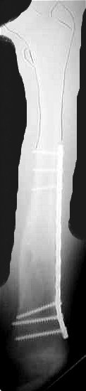
I've been using submuscular bridge plates inserted through small incisions, as have several others. Organized by Enes Kanlic, we have submitted an abstract with a number of these cases to the Academy meeting next year. It is really quite easy and reliable, and they heal like a house afire.
jeff Anglen
Date: Wed, 20 Jun 2001 21:32:24 EDT
From: OTS1
Jeff,
I have been following these plate cases with interest, as well as reviewing the photos and xrays. It is a great technique, but requires skill as a surgeon, and some of the reductions in comminuted fractures appear to have been a lot of work. While this technique is excellent in the right hands, a double locked nail is much easier, though I think the pq plates have a real place in the younger patient say 4-9? What do you think?
Roy Sanders
Date: Wed, 20 Jun 2001 22:42:15 -0500
From: Anglen, Jeffrey
Roy -
I agree for the most part. I will confess to having struggled a bit on the first few, but now I find it just as easy as a nail especially in the smaller kids. The reduction goals of length and alignment are similar, just bridging the comminution, and screw placement is the same as freehand interlock screw technique, or even easier as you can feel the plate holes with the drill bit and guide. I guess the nail is probably mechanically superior in the bigger kids.
Jeff
Jeffrey Anglen, MD FACS
Chief, Orthopaedic Trauma Service
Associate Professor, Orthopaedics
University of Missouri
Date: Thu, 21 Jun 2001 00:21:21 EDT
From: OTS1
Jeff,
I agree. See, the discussion about ender's and ex fix is probably moot, and i think ex fix is never really going to make it as a good solution in the U.S.
roy sanders
Date: Thu, 21 Jun 2001 11:32:35 +0600
From: Alexander Chelnokov Hello Roy,
Oac> I agree. See, the discussion about ender's and ex fix is probably moot, and i
think ex fix is never really going to make it as a good solution in the U.S.
Could you pls point me where i could learn more about the nailing
technique you mentioned? Otherwise i feel ex-fix is forever going to
make it as a good solution in Russia :-)
To be serious, actually other commonly available options here are bed
traction/open plating/open nailing through growth zones.
We've just started to implement closed nailing yet with conventional
nails available here. To date we've fixed 11 humeral shafts, 3 femurs and
2 forearms and at the moment i am happy with results. So your
experience will be very helpful.
Best regards,
Alexander N. Chelnokov
Date: Wed, 20 Jun 2001 23:19:13 -0700
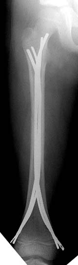
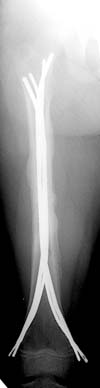
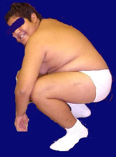
From: Carlo Bellabarba
alex,
here's a recent Ender example with three-month followup. sorry about the
quality of the injury image--there were better outside films that have since
disappeared. I still think this technique is useful, even in rotationally
unstable patterns such as this one. i take the nails out at 6 mos, and leave
them a little proud on purpose (this 10 year-old is a bit of chunkster so
they don't bother him) and suture them down so that they don't back out
more.
it aren't fancy, but it's quick and seems to work.
carlo bellabarba
Date: Thu, 21 Jun 2001 17:44:06 +0600
From: Alexander Chelnokov Hello Carlo,
I still think this technique is useful, even in rotationally
unstable patterns such as this one. i take the nails out at 6 mos, and leave
them a little proud on purpose (this 10 year-old is a bit of chunkster so
they don't bother him) and suture them down so that they don't back out
THX, i got the idea. I am also interested what nail Roy Sanders meant.
Best regards,
Alexander N. Chelnokov
Ural Scientific Institute of Traumatology and Orthopaedics
Date: Thu, 21 Jun 2001 14:32:03 -0400
From: Charles Mehlman
Two articles that I am aware of are:
Both of these articles relate to proximal femur growth abnormalities secondary
to IM rodding of pediatric femur fractures. Under the age of 13 years -
Gonzalez-Herranz found a 30% rate of bone abnormalities (such as coxa valga,
greater troch growth arrest, etc). Few seemed to require treatment, but they do
show a figure of a kid that underwent a corrective proximal femur osteotomy for
coxa valga. Raney et al spoke of "trochanteric epiphysiodesis" but were
uncertain of the functional disability from it.
I guess the point is, that even if you dodge the bullet of AVN - there are
still other snakes in the grass.
Jeff:
When are you taking plates out?
Charles T Mehlman, DO, MPH
Date: Thu, 21 Jun 2001 15:45:22 -0600
From: Kanlic, Enes M. M.D.
In deciding how to treat pediatric femur fractures the type of fracture and
weight of the patient are more important to be considered than the age.
"Problem" fractures (comminuted, long spiral, high sub or pertrochanteric,
supracondylar fx) do represent 43% of cases in Flynn, 2001 study, and 55% in
Heinrich,1994 study. For those fractures PERCUTANEOUS SUBMUSCULAR AND
EPIPERIOSTEAL BRIDGE PLATING is the best method.
Group of us (D. Smith, J. Anglen, S. Morgan, R. Pesantez, P.Cole) have done
more than 40 cases. Average OR time 103 minutes, x-ray time 105 seconds. No
infections or nonunions. All had excellent clinical result. Complications:
one temporary peroneal palsy, one valgus malalignment and one plate bending
after new injury (reduced closed and healed uneventfully).
Bridge plating provides elastic fixation for all type of fractures without
the need for bracing and casting, with excellent and reproducible results.
Plates are removed 6-12 month after injury.
In my opinion, stable fractures (transverse and short oblique shaft
fractures) should be treated with elastic intramedullary nails, what is even
less invasive than bridge plating.
Problems with other methods are:
1. Intramedullary elastic nailing: can not sufficiently stabilized "problem
fractures" (see above), in Flynn's study 70% of patients had to be put in
the cast; 9% did have complications.
2. External fixation: even after dynamization the refractering rate is to
high ( Minner, 2000: 21%; hardware support is simply to short). Bad scarring
and irritation - inflammation around pins are real problems (72% pins in
Miner's study were inflamed and required antibiotics). It is very difficult
if not impossible to treat supracondylar and subtrochanteric fractures with
this method.
3. Absolute stability osteosynthesis, compression plating: nonunion rate
is 10% in Fyodorov, 1999 study. To aggressive, "surgeon sensitive". 4. Locked, rigid intramedullary nailing: 5. Spica cast: for some authors is not desirable for patients heavier
than 50 lb (Stanitski, 1996).
I am attaching one of my cases.
Enes M. Kanlic, MD, PhD
Date: Thu, 21 Jun 2001 18:31:51 EDT
From: OTS1
Chuck,
as far as I know, the trochanteric epiphsiodesis is a risk, but is that a
problem in the adult? My sources as well as my reading suggest that it is not
a clinical problem in the adult in those who have developed it regardless of
etiology. Can you enlighten me?
roy
Roy Sanders, MD,
Date: Sun, 24 Jun 2001 12:16:31 -0400
From: Charles Mehlman
Roy:
There are at least two (2) papers addressing this topic...
(1) The first paper I know of that has addressed this is Ellen Raney's - She
(along with John Ogden & Dennis Grogan) reported five (5) patients that developed
increased neck valgus s/p IM nailing. One of their kids was nailed with a Rush
Rod - the others apparently with reamed IM nails. It is unclear from the paper
whether pirifromis fossa or tip of troch was starting point - but their figures
show Piriformis fossa site being used.
Their male patients were between 11 and 13 years of age and their female
patients were 9 and 11 years of age. Within the context of two to seven year
follow-up - NO FUNCTIONAL PROBLEMS WERE IDENTIFIED.
(2) The second paper to address this is Gonzalez-Herranz's for the British
JBJS. This group of Spanish authors studied IM nailing effects on 34 chidlren
with an average age of about 10 years. Twenty-two (22) kids were treated for
femur fractures and 12 were treated for femur tumors. Coxa valga and decresed
troch height were among the "things" they saw. Their worst case looks like it
was 6-year-old nailed with a K-nail thru the piriformis fossa that developed coxa
valga bad enough to require a corrective osteotomy at age 18.
Take home points in my mind: (1) there seems to be little price to pay
regarding insulting the trochanter in adolescents, and (2) antegrade nailing in
the less than 10 crowd is probably not a good idea at all.
Charles T Mehlman, DO, MPH
Date: Sun, 24 Jun 2001 22:25:25 EDT
From: OTS1
Chuck,
I don't disagree with you. I think a real study needs to be done. Clearly if
you go in through the piriformis and don't get the vessels, you can still
affect growth. Also if the capital physis has not separated from the
trochanteric apophysis, this can be a problem, hence the younger child is
still a problem. The question is: does nailing through the troch apophysis
once separated from the capital epiphysis affect the neck angle, the
abductors, the patient's gait and or function. I plan to find out, because
traction, spica casts, ender nails, bridge plating, and/or ex fix's are not
the answer to a long bone fracture.
thanks for reviweing the literature.
roy sanders
Date: Mon, 25 Jun 2001 06:36:40 -0500
From: Anglen, Jeffrey
When are you taking plates out?
6-12 months, when there seems to be solid healing and when it is convenient
for the family. I let them go right back to full WB on the leg, but try to
restrict sports, climbing, skating, etc. for a month or 6 weeks. So far (
knock on wood) no re-fractures.
Jeff Anglen
Jeffrey Anglen, MD FACS
Date: Mon, 25 Jun 2001 12:25:28 -0400
From: William Obremsky
What is the rationale that you tell the family for plate removal? Why do we
treat femur fxs differently than BB forearm fxs? I have also removed the plates
or flexible IM rods, but do not have a really good explanation to the family and
have begun to present it as an elective procedure that the parents may decide to
pursue.
Bill Obremskey
Date: Mon, 25 Jun 2001 19:50:55 -0500
From: Anglen, Jeffrey
Bill -
To tell you the truth, I've never had a family question my recommendation
for plate removal. I remove them because they have so much growing to do
that I am afraid they will become encased in bone and be impossible to
remove later in the situation of another fx or desire to join the army, or
whatever. Usually we don't expect the forearms we plate to double or triple
in size through growth. I don't know for sure that having a plate
completely surrounded by bone would be a problem outside of those perhaps
unlikely situations, but it makes me nervous. It would actually be
interesting to see what it looks like at adulthood.
Jeff
Date: Mon, 25 Jun 2001 20:03:42 -0500
From: Steven Rabin
I think we remove the plates for the reasons Jeff gives. If the bone
surrounds them and we have to remove them at a later date - - then the removal is difficult and potentially complicated by the risk
of refracture. I recently removed a tibial plate encased by bone - the patient's
father, and three uncles had all had amputations from diabetes-related infections,
and the patient wanted it out before it became a potential problem (he wasn't
diabetic yet). It was a big operation, and although I didn't break the bone
taking it out, I could have. Taking them out early is a lot less destructive
and traumatic than taking them out late. I realize that the majority of patients will probably never be bothered
by the plate, but I think they should at least understand the options.
Date: Tue, 26 Jun 2001 09:30:18 +0600
From: Alexander Chelnokov
Hello Steven,
SR> I think we remove the plates for the reasons Jeff gives. If the
bone surrounds them and we have to remove them at a later date -
Doesn't it mean that methods which don't require a separate surgery
for hardware removal (which sometimes is more complicated than initial
surgery) have some advantages?
Best regards,
Date: Mon, 25 Jun 2001 22:52:25 -0500
From: Steven Rabin
Hello Alexander,
Yes, options that would not require hardware removal have definite advantages
because they avoid the risks and expense and hassle of the second surgery. But,
as discussed by many already, I think external fixators have too high a risk of
refracture. Bone can overgrow the ends of flexible nails and with further growth
they can get lost in the medullary canal making it extremely difficult to
retrieve them at a later time. Locked rods such as the Trigen system may be
easier to find and retrieve later even if bone overgrows them since they'll stay
locked in the same place, but have the issues discussed concerning violating the
greater trochanteric apophysis.
There's no perfect solution yet. If it were my child, I'd rather have him/her
undergo a hardware removal, than suffer a refracture.
Date: Tue, 26 Jun 2001 18:07:21 +0600
From: Alexander Chelnokov
Hello Steven,
SR> think external fixators have too high a risk of refracture.
Maybe the feature is not a result of the method in whole, but only of
some aspect of its implementation, which can be revealed and
eliminated?
[...]
SR> There's no perfect solution yet.
God bless the situation! :-)) It leaves us place and role to balance
all advantages and disadvantages which can't be expressed as simple
numeric values otherwise human surgeons soon would be replaced by
computer-assisted devices.
SR> If it were my child, I'd rather have him/her undergo a hardware
SR> removal, than suffer a refracture.
Remembering some disastrous removals and that re-fractures usually
heal very quickly and easy, i wouldn't be so categorical...
Let our children be healthy though :-)
Best regards,
Date: Wed, 27 Jun 2001 15:36:10 -0400
From: William Obremsky
Jeff,
I would agree w/ you and try to explain these issues to families and have
routinely removed FIMN or plates, but I always like to jusify in my own mind the
rationale or risk/benefit ratio of procedures. The unknown question is: what %
of kids that have internal fixation would have a second similar fx or develop
ipsilateral arthritis that would require an arthroplasty where the implant from
the fx would interfere w/ the arthroplasty? Refracture around an intact implant
is unlikely and who knows what the answer to degenerative problems will be when
these pre-teens become aging adults? We were hardly doing arthroplasty 40 years
ago. Is the probably small percentage and probability of a problem of retained
hardware worth the risks and costs of 100% hardware removal? Sounds like a cost
/benefit analysis by an economist is needed
Bill Obremskey
Date: Fri, 6 Jul 2001 18:46:53 +0600
From: Alexander Chelnokov
Hello Steven,
SR> Yes, options that would not require hardware removal have definite
advantages because they avoid the risks and expense and hassle of
I've just found IMHO a good systematic view at
http://www.aaos.org/wordhtml/anmt2001/sympos/sympc.pdf
===============================
OPERATIVE MANAGEMENT OF PEDIATRIC FEMUR FRACTURES Paul D. Sponseller, MD; Baltimore, MD
I. General Principles A. Indications - Age and surgeon experience Other relative indications for surgery B. Type of Operative Fixation - Preferences by Age - reamed nail through piriformis II. Specific Operative Techniques A. External Fixation External Fixator Results (Blasier, Turski & Aronson JPO 1997) Refracture after External Fixation B. Plate Fixation (Results -Ward, Sturm) C. Intramedullary Rods - advantages - Disadvantages - Avascular Necrosis ? Caused by injury to ascending cervical artery (Huurman 1996) 1. Flexible Nails -Technique: flexible nails -Flexible nails: aftercare -Complications of flexible nails: 2. Rigid Nails III. Cost Factors Charge Comparison (Stans, Morrissy 1997) Charge Comparison-Conclusions IV. Conclusion References: Aronson J Tursky A: External Fixation of Femur Fractures in Children.
J. Pediatr. Orthop 12: 157-163, 1992 Blasier RD, Aronson J, Tursky EA: External
Fixation of Pediatric Femur Fractures. J Pediatr. Orthop 17: 324-246, 1997. Fyodorov I, Sturm PR, Robertson WW: Compression-plate fixation of femoral
shaft fractures in children aged 8-12 years. J. Pediatr. Orthop. 1999, 19:
578-581 Huurman W: Avascular Necrosis After Intramedullary nailing of Pediatric
Femur Fractures. Presented at the annual meeting of the Pediatric Orthopaedic
Society of North America, 1996. Mamberger N, Stevens P, Smith J, Santora S, Scott S, Anderson J: Intramedullary
nailing of femoral fractures in adolescents. J. Pediatr. Orthop 20(4): 482-484,
2000. Mendelow MJ, Anastasios D, Kanellopoulos AS, Mencio GA, GreenNE: External
Fixation of Pediatric Femur Fractures. Orthop Trans1997;21: 185 Miner T, Carroll KL: Outcomes of External Fixation of Pediatric Femoral
shaft fractures. J. Pediatr. Orthop 20(3): 405-410, 2000. Skaggs DL, Leet A, Money MD, Shaw BA, Hale JM, Tolo VT: Secondary fractures
associated with exterhal fixation in pediatric femur fractures. J. Pediatr.
Orthop 1999: 582-586 Sola J, Schoenecker PL, Gordon JE: External Fixation of Femoral Shaft
Fractures in Children. Enhanced stability with user of an auxiliary pin.
J. Pediatr. Orthop 19(5): 587-591, 1999 Tolo VT: Treatment of Fractures of the long bones and pelvis in children
who have sustained multiple injuries. J. Bone Joint Surg 82(A): 272-280,
2000 Vransky P, Bourdelat D, Al Faour A: Flexible Stable Intramedullary Pinning
Technique in the Treatment of Pediatric Fractures. J. Pediatr. Orthop 20(1):
23-27, 2000 Ward WT, Levy J, Kaye A: Compression Plating for child and adolescent
femur fractures. J. Pediatr. Orthop 12: 626-632, 1992 ===============================
Best regards,
Alexander N. Chelnokov
Ural Scientific Institute of Traumatology and Orthopaedics
str.Bankovsky, 7. Ekaterinburg 620014 Russia

seattle
str.Bankovsky, 7. Ekaterinburg 620014 Russia
Assistant Professor Pediatric Orthopaedic Surgery
Division of Pediatric Orthopaedic Surgery
Children's Hospital Medical Center Cincinnati
For Adam Starr: Synthes titanium elastic nails offer 5 different diameters
(2.O - 4.0 mm), easy length adjustment and excellent instrumentation.

TTUHSC Ortho Dept
El Paso, Texas
Tampa, Florida
Assistant Professor Pediatric Orthopaedic Surgery
Division of Pediatric Orthopaedic Surgery
Children's Hospital Medical Center Cincinnati
Chief, Orthopaedic Trauma Service
Associate Professor, Orthopaedics
University of Missouri
UNC Orthopedics
Alexander N. Chelnokov
Ural Scientific Institute of Traumatology and Orthopaedics
str.Bankovsky, 7. Ekaterinburg 620014 Russia
Alexander N. Chelnokov
Ural Scientific Institute of Traumatology and Orthopaedics
str.Bankovsky, 7. Ekaterinburg 620014 Russia
Ural Scientific Institute of Traumatology and Orthopaedics
str.Bankovsky, 7. Ekaterinburg 620014 Russia