
Date: Tue, 24 Apr 2001 14:20:11 +0200
From: J.C. Goslings
Subject: Heterotopic ossification??
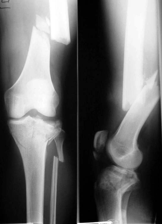
This 44 year old male was brougth in after a MVA. He was respiratory and hemodynamically stable, no neurological defecit. After assessment according to the ATLS protocol and subsequent evaluation the following injuries were diagnosed:
The aortic disruption was treated immediately by placement of an endovascular stent. The wounds were debrided and irrigated, the fractures were bridged by external fixation or splinted.
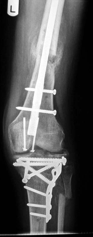
His general condition improved and he returned to the ward after 5 days at the intensive care unit. In the next 2 weeks all the fractures were subsequently treated by internal fixation. In the left femur retrograde unreamed nailing was performed. Two screws were placed percutaneously in the patella. The tibial plateau fracture was treated by plate and screw osteosynthesis under fluoroscopic control.
|
|
|
Post-operatively, the knee was immediately put on a CPM machine. Despite our functional treatment 2 months after the accident his knee flexion is limited to 70 degrees; knee extension is normal. It is our impression that the flexion is limited by the (palpable) ossification around the femoral fracture. What are your suggestions about further treatment of this problem and how would you improve the knee function?
Thank you; in advance,
Date: Wed, 25 Apr 2001 12:38:04 -0500
From: Steven Rabin
I'd like current x-rays to give a definite opinion, but.. At 2 months, it may not be healed enough for further surgery, and further PT and rehab may still improve function enough to avoid further surgery. When the fracture heals, and the abundant excessive callus becomes mature, then excision of the excess bone with possible Judet quadricepsplasty, and possible lysis of intra-articular adhesions might restore functional motion.
Date: Wed, 25 Apr 2001 16:00:03 -0400
From: J. Tracy Watson
Once Fx has healed... check CT scan to determine orientation of HO...including whether of not it has enveloped the medial vasculature.....then excision HO with Probable Judet quadricepts plasty...(a big operation ).
JTW