

Date: Thu, 11 Dec 2003 15:28:53 -0500
Subject: Acetabular impaction/fracture
I am presenting this case for opinion of the list members.
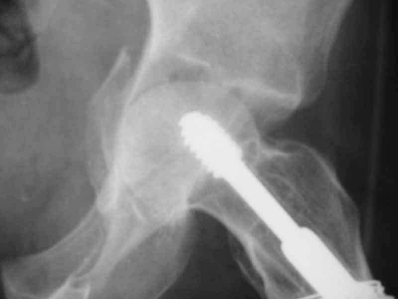
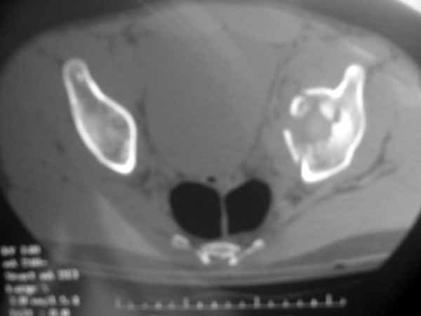
39 yo gentleman (previously treated by referring surgeon for proximal femur fx with screw and side place device), s/p fall onto his left lateral proximal femur while bicycling. Plain films and CT demonstrate significant anteromedial impaction injury to acetabular dome and medial displacement of femoral head/quadrilateral plate. "Gull Sign" noted on all AP pelvis and Judet's. I appreciate the difficulty in obtaining and maintaining the reduction (Anglen et al, JOT, October, 2003, 625-634).
Thanks for all comments and input.
|
|
|
|
Robert A. Hymes, MD
INOVA Fairfax Hospital
Fairfax, VA
Date: Thu, 11 Dec 2003 16:30:08 -0600
From: Anglen, Jeffrey
Yipes. Because of his young age, I would suggest to him that we attempt reduction and fixation - going through an ilioinguinal approach, and making sure that any fixation screws were well away from the joint - enough to not interfere with subsequent arthroplasty. I would tell him about the difficulty anticipated with reducing and stabilizing that central impaction, and the implications for later arthroplasty. Nonetheless, if you can reduce and fix that anterior column, you may be able to get the head to stay under the remaining dome. I think it is worth a shot in this age patient. If he was 69, I'd opt for non-operative treatment until the proximal femur was healed, then arthroplasty as indicated by the symptoms.
I have not been very successful with over-the-rim spring plates, wiring, or buttressing of the quadrilateral plate as has been written about in the literature.
Jeff
Jeffrey O. Anglen MD FACS
Boone Orthopaedic Associates
Clinical Professor of Orthopaedics
University of Missouri
Date: Thu, 11 Dec 2003 14:29:43 -0800
From: Chip Routt
Impaction fractures can be managed with reduction and fixation.
Enclosed is a similar impaction example on our patient's left side.
 |
 |
 |
The ilioinguinal exposure is good. Impaction fragment reduction and support bone grafting can be done either thru the primary fracture prior to its reduction, or thru an iliac fossa cortical window...depends on the fracture details. We used a cortical window for this patient. He's a heavy equiptment mechanic now 18 mos after injury.
It's difficult, but good to do-
Chip
M.L. Chip Routt, Jr.,M.D.
Professor-Orthopedic Surgery
Harborview Medical Center
Seattle, WA 98104-2499
Date: Thu, 11 Dec 2003 18:29:39 -0700
From: John T. Ruth
Chip, how do you judge the adequacy of your reduction using a cortical window? Intraop. imaging? Visualization through the fracture site?
Date: Sat, 13 Dec 2003 10:41:19 -0500
From: Jason Nascone, M.D.
Bob
I'd vote for ORIF via Ilioingiunal approach. Its usually a blind reduction that I judge on image. Lamina spreader in the fracture and a cobb to reduce the impaction while pulling lateral traction on a schantz pin in the prox femur and graft the stink out of it to hopefully reduce the increased volume caused by the impaction. If you can't reduce the volume then he's going to want to displacing medially
Good luck
Jason W. Nascone, M.D.
Assistant Professor
University of Maryland Department of Orthopaedics
RA Cowley Shock Trauma Center
22 South Greene St.
Baltimore, MD 21201
Date: Mon, 15 Dec 2003 11:48:39 -0800
From: Chip Routt
JR-
If the primary fracture site is still open and the head is beneath the intact dome segment, the impaction fragment(s) is(are) reduced to the head and grafted from within the primary fracture site and/or thru a cortical window, and the reduction and grafting are assessed by direct visualization and also intraop fluoro...then the primary fracture is reduced and secured.
If the primary fracture site is initially reduced and secured, then the impaction fragment is reduced with an impactor thru a strategically located cortical window and bone grafted...in this situation, the reduction must be assessed by multiplanar fluoro (or intraop CT if you're really fancy!).
It's best to hold a bit of distraction on the head as the dome fragment is tamped into place, then tamped an additional 1-2 mm...as the head distraction is then relaxed, the " slightly over-reduced" impaction fragment settles perfectly.
The window location sorts out in the preop plan.
Chip
M.L. Chip Routt, Jr.,M.D.
Professor-Orthopedic Surgery
Harborview Medical Center
Seattle, WA 98104-2499