
Date: Mon, 22 Apr 2002 17:57:51 -0400
Subject: Tibia Varus IR malunion
Advice on this patient?
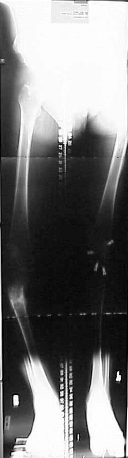
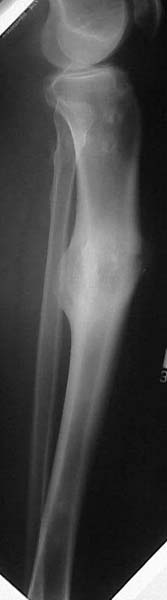
49 yo college professor s/p right proximal tibia fx w/ compartment syndrome. Pt had fasciotomies, ex-fix and delayed percutaneous plating laterally w/ LISS. He had late deep infection at 6 mos and plate removed and fx appeared healed at time. Pt had some varus and deformity, but has collapsed further w/ 25 degrees varus, 15 degrees recurvatum and 20 degrees IR deformity. Lateral wound is healed. ESR and WBCs are normal, 9 mos post injury. Deformity is cosmetically and functionally not acceptable.
|
|
|
Votes for:
1) 3 plane correctional osteotomy per Sangeorzan
2) opening wedge medial transverse osteotomy, 90 degree humeral blade fixation and graft
3) transverse osteotomy, medial opening wedge and IMN correction and grafting
4) any others?
Thanks for any input.
Bill Obremskey
UNC Dept of Orthopedics
Date: Mon, 22 Apr 2002 16:28:48 -0700
From: Bruce Sangeorzan
bill
the malunion (nonunion? hard to tell) is ideally suited to a single cut osteotomy.
bruce
Date: Mon, 22 Apr 2002 16:40:02 -0700
From: John Ruth
Bill
Based on the AP, measurements you stated and the history of infection, I would recommend a medial opening corticocomy or dome type osteotomy done through small incisions and Ilizarov correction. I am sure he is short and this would help restore some length.
Date: Mon, 22 Apr 2002 18:44:48 -0600
From: Thomas Higgins
Working off of John's idea of Ilizarov in consideration of infectious hx and possible LLD, this may be addressed nicely with a spatial frame. It offers the Ilizarov option with the ability to more easily facilitate a multiplanar correction.
Date: Tue, 23 Apr 2002 07:24:15 -0400
From: Kevin Pugh
This is ideally suited for an Ilizarov type correction. The short peri-articular fragments can be purchased, the deformity corrected, hardware avoided at the site of previous infection, potentially bad soft tissues treated well, and length obtained (from this site or at another level).
Kevin J. Pugh, MD
Chief, Division of Trauma
Department of Orthopaedics
The Ohio State University
Columbus, OH 43210
Date: Tue, 23 Apr 2002 17:53:22 +0600
From: Alexander Chelnokov
Hello William,
WO> removed and fx appeared healed at time. Pt had some varus and deformity, but has collapsed further w/ 25 degrees varus, 15 degrees recurvatum and 20 degrees IR deformity. Lateral wound is healed. ESR
An ideal case for Ilizarov-type correction with closed corticotomy and ring or hybrid frame with hinges. Some olive wires into the proximal fragment, two 6 mm titanium threaeded pins to the distal. I would correct angular deformity first, then re-estimate rotation and if found proceed to its gradual correction.
Best regards,
Alexander N. Chelnokov
Ural Scientific Institute of Traumatology and Orthopaedics
str.Bankovsky, 7. Ekaterinburg 620014 Russia
Date: Tue, 23 Apr 2002 14:12:36 +0100
From: Nuno Craveiro Lopes
William,
You can see at http://clientes.netvisao.pt/nfrancac/preop01.htm how we simultaneously correct shortening and axial deviation with a Ilizarov frame.
Best regards,
Date: Tue, 23 Apr 2002 09:20:44 -0400
From: Kevin Pugh
Bill,
I just went back and looked at the films again. You have at a minimum a 4 axis deformity (3 angulations and length). An Ilizarov will be somewhat complicated. I would do this with a Spatial frame to allow simulatneous correction of all deformity with one frame. The "prebuild" is easy, and the hinge is virtual.
KP
Date: Tue, 07 May 2002 06:42:21 -0400
From: William Obremsky
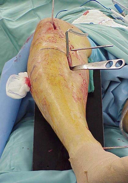
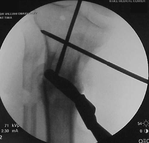
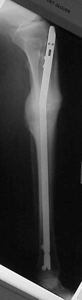
Thanks for the input on the tibia malunion w/ multiple plane deformity. I decided on a single cut osteotomy and IMN fixation based on desire to walk early, return to professor duties, and my previous experience w/ similar deformities (see another tibial malunion -below). The opening wedge was packed w/ a combination of autograft, allograft and demineralized bone.
 |
 |
 |
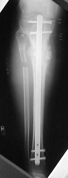 |
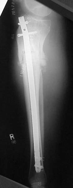 |
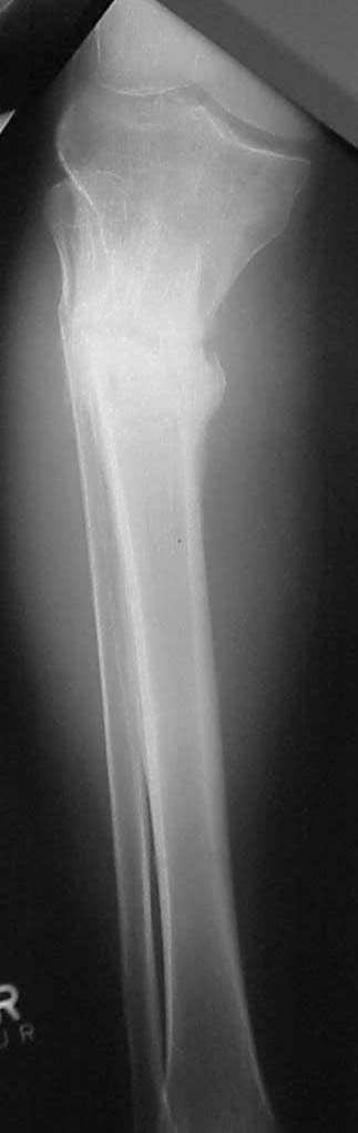
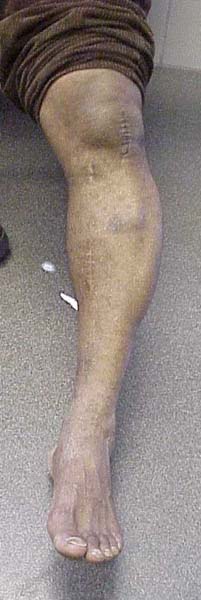
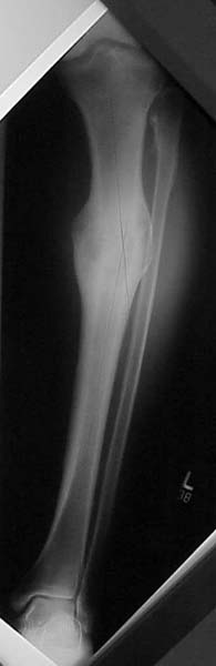
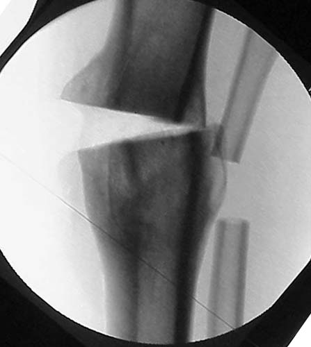
Another tibial malunion -
Pt was 10 yrs s/p open tibia fx treated in a cast w/ IR, varus, 2 cm short, and recurvatum. He has done well post op.
 |
 |
 |
 |
 |
 |
Bill Obremskey
Date: Tue, 07 May 2002 12:05:04 -0400
From: James Carr
Nice!
jbc
Date: Sat, 11 May 2002 10:30:26 +0600
From: Alexander Chelnokov
Hello William,
WO>The opening wedge was packed w/ a combination of autograft, allograft and demineralized bone.
Is there any experience with a bit different approach - closed corticotomy, 7-10 days of correction by an XF with hinges with formation of wedge-shape regenerate, and then early conversion to closed IM nail?
It would require no grafting and much less surgical exposure. Disability and decrease of quality of life due to a couple of weeks in XF looks comparable if not less to that with the open surgery with graft harvestng. Intuitively it also must be of less risk of local complications. On the other hand, it is two surgical procedures (though less invasive each), two anesthesiae...
Best regards,
Alexander N. Chelnokov
Ural Scientific Institute of Traumatology and Orthopaedics
str. Bankovsky, 7. Ekaterinburg 620014 Russia
Date: Sat, 11 May 2002 17:51:58 EDT
From: Tadabq
Alex has proposed correction of tibia shaft angulatory malunion by ring fixator, corticotomy and opening "wedge" followed by IM nail.
Benefits vs opening wedge osteotomy:
1. No need for bone graft
2. Gradual correction (perhaps more precise)
3. Minimal dissection at the correction site
4. Nail fixation for functional aftercare
Drawbacks
1. Two operations
2. Risk of infection of nail after XF
3. Expense of ring fixator AND nail
4. Expertise in both ring XF and nail required
5. Would regenerate bone hold up during and after nailing?
TD
Date: Sun, 12 May 2002 10:20:09 +0600
From: Alexander Chelnokov
Hello Tom,
THX for the balanced list of pro and contra.
Tac> Drawbacks
Tac> 2. Risk of infection of nail after XF
As reported by many colleagues early conversion has very low risk.
Tac> 3. Expense of ring fixator AND nail
Fixator comes back in a couple of weeks except wires/pins.
Tac> 4. Expertise in both ring XF and nail required
IMHO many surgeons have both.
Tac> 5. Would regenerate bone hold up during and after nailing?
To preserve regenerate during nailing it should be performed with fixator in place, just wires/pins should be removed except last proximal and distal, to keep length/axis/rotation.
Best regards,
Alexander N. Chelnokov
Ural Scientific Institute of Traumatology and Orthopaedics
str. Bankovsky, 7. Ekaterinburg 620014 Russia