
Date: Friday, February 19, 1999 10:04 PM
Subject: Talus fracture
Dear all,
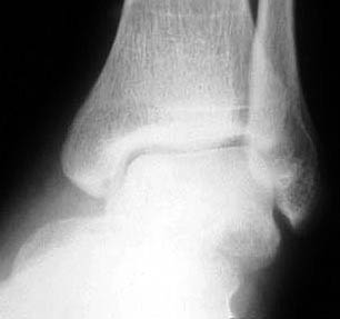
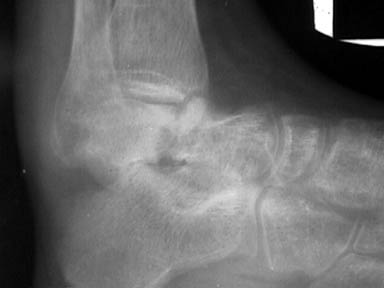
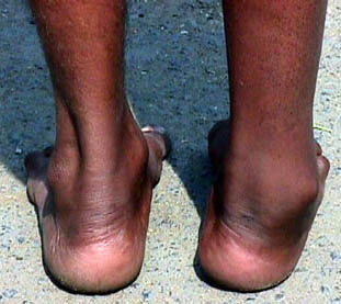
Am seeking advice on management of 3 months old displaced and ununited fracture neck of Talus. Patient is 35 years old manual worker. At present he cannot weightbear because of pain and his heel is going into varus. His clinical picture, initial xrays and present xray are attached.
TIA - Rajat Varma FRCS, Indore, India.
| Injury Films | |
|
| 3 months | |
|
Date: Fri, 19 Feb 1999 15:47:14 -0600
My vote would be for a Blair tibiotalar arthrodesis. Discard the dome, roughen up the anterior tibia and put the talar neck right against it, fix with 2 screws from the back of the tibia into the talar head. This has worked better for me than the business with sliding the anterior tibial cortex down, etc.
Jeff Anglen, MD , Chief, Orthopaedic Trauma Service, University of Missouri
Date: Fri, 19 Feb 1999 16:17:44 -0700
Chronic nonunion of talar neck in 35 year old laborer with moderate deformity. Xrays nicely presented. I doubt reduction and fixation of the talar neck fracture would be successful. I would consider reduction of the foot in relationship to the knee and leg and probable arthrodesis of the talus body to the tiba and calcaneous; perhaps with a retrograde nail from Smith & Nephew.
Tom DeCoster
Date: 19 Feb 99 17:46:07 -0800
Dr Rajat
This is an interesting and challenging case. He hs 2 problems, position and pain. I assume he still has ankle motion. You would serve him well to preserve as much of his motion as you can with priority given to the ankle joint. I suggest CT to best understand the deformity. You will find that the body component is plantar flexed relative to the rest of the foot. The neck fragment appears viable. it would be helpful to know its relationship to the navicular and calcaneus. This could be seen from a modified ap view of the foot and/or from the CT scan. Restoring the position of the body piece is challenging and will require a femoral distractor from the calcaneus to the talus. you will need to elevate the front of the body piece. You will need bone graft and minifrag plates to maintain the alignment of the talus, IF you cannot maintain the position of the subtalar joint, I suggest you arthrodese the subtalar joint. Do all you can to preserve the ankle.
bruce
Bruce J Sangeorzan MD, Professor Orthopedic Surgery, U of Washington
Date: Sat, 20 Feb 1999 22:53:39 +0530
Dear Dr. Rajat,
According to Hawkin's classification your patient seems to have type II fracture neck talus with subluxation or dislocation of the subtalar joint, compromising the two blood supply sources and thus resulting in a higher incidence of avascular necrosis .
Moreover x-rays do not show Hawkin's Sign which is usually visible in 6-12 weeks after injury , thus going more in favour of avascular necrosis.So better to go for bone scanning first and to the final treatment to be done accordingly. Please let the list know how did you manage the case?
DR. M. P. SHRIVASATAVA, CONSULTANT ORTHOPAEDIC SURGEON, SIDDHARTHA APOLLO HOSPITAL, KATHMANDU, NEPAL.
Date: 20 Feb 99 20:49:26
Hi
I have 2 similar cases finished and third in progress treated with tibiocalcaneal fusion and tibia lengthening. A couple of months ago i sent images with an example of this approach to Randale Sechrest, maybe he didn't erase the x-rays and could kindly upload them to his site. Tibiotalar andtalocalcaneal fusion in such cases IMHO can take more time to heal.
 |
 |
 |
 |
Best regards, Alexander N. Chelnokov, Ural Scientific Institute of Traumatology and Orthopaedics, str.Bankovsky, 7. Ekaterinburg 620014 Russia
Date: Tue, 23 Feb 1999 08:59:56 +0530
Thank you all for your suggestions.
This patient has a stiff ankle as well. So I plan to do tibiotalarcalcaneal fusion as has been suggested. However I have no experience with retrograde nail in this situation and am planning to do antegrade nail from tibia into calcaneum . Any tips or references would be welcome.
TIA - Rajat Varma FRCS, Indore, India.
Date: Tue, 23 Feb 1999 09:19:39 -0500
From: Kevin Pugh
A retrograde nail would be easier, and probably more accurate at getting the joints in the position you would like. Your Richard's rep, or the rep of the company whose nail you plan to use, would have access to a technique.
Date: 24 Feb 99 21:41:20 -0800
As a foot/ankle surgeon who wears a trauma hat, I cant pass this discussion of tibiocalcaneal fusions without reminding everyone that this is a salvage procedure. please dont sacrifice the ankle and subtalar fusion without putting up a fight. Pantalar function is not very good. patient satisfaction is low. people are only happy when the alternative is subtantial pain. a Pantalar fusion creates a living prosthetic limb without the potential for energy return.
Bruce Sangeorzan
Date: Thu, 25 Feb 1999 23:14:49 +0500
>as a foot/ankle surgeon who wears a trauma hat, I cant pass this discussionof tibiocalcaneal fusions without >reminding everyone that this is a salvageprocedure. please dont sacrifice the ankle and subtalar fusion without puttingup a fight.
What approach should be recommended in comminuted cases? Especially in delayedadmission? In fractures with primary bone defect of talus? When and in what casesdecision about fusion should be considered?
> Pantalar function is not very good. patient satisfaction is low.
Comparatively to what?
>people are only happy when the alternative is subtantial pain. a Pantalarfusion creates a living prosthetic limb without the potential for energyreturn
How many cases after reconstruction in severe talar fractures sooner or laterrequire the fusion?
Best regards, Alexander N. Chelnokov, Ural Scientific Institute ofTraumatology and Orthopaedics, 7, Bankovsky str. Ekaterinburg 620014 Russia
Date: Fri, 26 Feb 1999 14:29:54 -0500
Dear Dr. Rajat:
I am Dr. Raymond Sullivan, the Foot and Ankle specialist at UCONN. I think in seeing this briefly the patient would best be treated with ORIF and tricortical Iliac crest bone graft. The lager portion of the graft would be placed plantar an medial to help stabilize the reconstruction. The screws should be placed from posterior into the talar head. Of course the patient should know about the > 50% incidence of post-op AVN of the body.
Date: Fri, 26 Feb 1999 17:34:50 EST
Bruce states the key point here, in fact it sounds like something I wouldsay,..... see occasionally Tampa and Seattle think alike!
Roy Sanders, M.D., Tampa, Florida
Date: Sun, 28 Feb 1999 10:21:13 -0700
Reply-To: Alexander Chelnokov
This case appears to be an tibio-calcaneal fusion (?after talus fractureand osteonecrosis?) with lengthening. It appears you used two sites forcorticotomy both diaphyseal? Is this correct and was the purpose tospeed the process? It appears you have made oblique or step cuts of thefibula. Is this correct and is the purpose to enhance regenerate? Whatis your current opinion regarding the importance of preservation of themedullary vasculature in distraction osteogenesis?
Tom DeCoster
Hi All especially Tom
This case appears to be an tibio-calcaneal fusion (?after talus fractureand osteonecrosis?) with lengthening.
More or less so. It was a case of delayed admission (more than 2 month afterinitial injury when in multiple trauma the talus fracture stayed untreated).So when the talus was exposed there was a shapeless conglomerate. It wasmuch easier to remove it.
It appears you used two sites for corticotomy both diaphyseal?
Yes. Interestingly that before the corticotomy i performed a failed attemptto grow a regenerate at the site of tibiocalcaneal contact as well as inknee arthrodesis with lenghthening, after distal femur osteoblastoclastomaremoval. I started tension at 10th day postop with usual 1 mm/day, x-ray at10 th day of tension showed no movement 8-[ ] Tibia and calcaneus alreadywas united! So the corticotomy was performed. Hardware was removed at 115day after corticotomy. Total 137 days with apparatus.
Is this correct and was the purpose to speed the process?
Yes.
It appears you have made oblique or step cuts of the fibula. Is thiscorrect and is the purpose to enhance regenerate?
No. Generally in tibial lengthening regenerate of fibula is of no care.Oblique oteotomy is IMHO safer and more convenient to perform when smallincision is used.
What is your current opinion regarding the importance of preservation ofthe medullary vasculature in distraction osteogenesis?
We of course try to preserve medullary space but [whisper not to be heardby orthodoxal Ilizarovians] in cases when my chisel definitely penetratedinto the medullary canal, regenerates of usual quality were then occuredtoo. IMHO periosteal blood supply plays leading role to provide good qualityregenerate. So one should avoid soft tissue stripping first. If hepreserved the canal too - good for him, if not - nothing to be afraid of.
Best regards, Alexander N. Chelnokov, Ural Scientific Institute of Traumatologyand Orthopaedics 7, Bankovsky str. Ekaterinburg 620014 Russia