Date: Wed, 18 Apr 2001 21:24:05 +0530
Subject: Pelvic fracture
From: Dr. M. P. Shrivastava
Dear Colleagues,
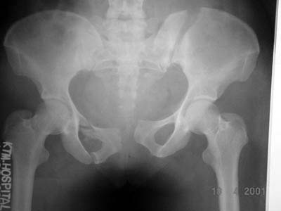
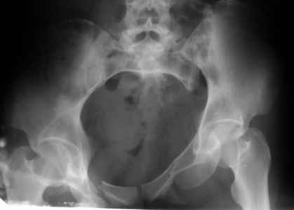
A female patient aged 40 years sustained pelvis fracture at different levels about a week ago. Kindly give your valuable opinions how to treat this difficult fracture? Image is attached herewith
Dr. M. P. Shrivastava
Consultant Orthopaedic Surgeon
Nepal Medical College Teaching Hospital
Jorpati, Kathmandu, Nepal
|
From: Yehia Basyoni,MD.
Sent: Wed, 18 Apr 2001 21:24:05 +0530
Dear sir
I think the proper way to manage is to fix the posterior iliac fracture with anterior plating, one on the iliac crest and another anteriorly. The anterior fracture will be approached through LT ilioinguinal approach extending to RT superior pubic ramus and fix with a reconstruction plate extending from RT iliac bone to LT pubic bone
Sincerely
Yehia Basyoni,MD.
Ass. Prof in Orthop.
Mansoura University
Egypt.
From: Cihangir Tetik M.D.
Sent: Wed, 18 Apr 2001 21:24:05 +0530
Dear Shrivastava
External fixator is the best way for treatment of this fracture. Three Shanz screws for each iliac wing longitudinally is necessary and then something to connect these pins are enough to secure the reduction. Anatomic reduction is prefered but not necessarily. Many external fixator systems you can find or you can customize.
Good luck
Cihangir Tetik M.D.
Date: Thu, 19 Apr 2001 16:30:44 -0700
From: John Ruth
ORIF posterior iliac wing fracture with 6.5 lag screw between the tables of the ilium. Possible iliosacral screw depending on whether or not the SI joint is involved (can't tell from solitary AP view of pelvis). Also would do open plating of symphysis pubis disruption.
Date: Thu, 19 Apr 2001 18:55:52 -0500
From: Kyle Dickson, MD
This seems like an unstable injury therefore an external fixator is inadequate fixation (numerous papers show this). Anterior plating is difficult unless you cross the si joint. Furthermore, anterior reduction is more difficult because it is difficult to use clamps to hold the reduction while you assess the reduction. Therefore I would plate it posteriorly and use an iliosacral screw if the si joint is unstable (doubt it) and then plate the symphysis anteriorly.
kyle
Date: Fri, 20 Apr 2001 07:57:50 -0700
From: Thomas A. DeCoster
This 40 year old woman in Turkey is a week after a pelvic ring disruption. Her injuries include a fracture of the left ilium posteriorly with displacement, a pubic symphysis disruption with about 5 cm of displacement and right superior pubic ramus and ischial ramus fractures. The left ilium fracture extends from the pelvic brim to the iliac crest and appears to begin anteriorly near the sacro-iliac joint.
These patients typically suffer from 3 problems. Some have life threatening hemorrhage initially. An external fixator has been very useful in this setting. She's over a week post injury and this does not seem to be a problem in this patient.
They are unable to walk or even move around in bed comfortably for many months after injury. A variety of techniques may be helpful at lessening this problem. Her injury pattern is not easily fixed with any technique. I might use an external fixator for this problem as the best benefit to risk ratio of the fixation alternatives.
They have chronic pain and loss of function. Anatomic reduction and stable fixation to healing is appealing to try to minimize this problem. Some people would recommend anatomic reduction and fixation of the left posterior ilium, the symphysis, the right superior pubic ramus and probably even the ishical ramus fractures.
Others have suggested fixation of the posterior ilium by posterior approach and lag screw or plate. That can be hard but not impossible to combine with ORIF of the anterior ring. Approaching the posterior ilium fracture from anterior would require fixation to the sacrum (because there is so little (or none) ilium medial to the fracture anteriorly) which is never perfectly secure; but has the benefit of being easier to combine with anterior ring surgical approach.
Some have suggested reduction and fixation of the symphysis (presumably without fixation of the right superior pubic ramus) and others have suggested fixation of the symphysis and the superior pubic ramus that would require extending the dissection to the right ilium proximal to the right acetabulum. (ilioinguinal approach).
That is a big dissection but would be the technique most likely to produce the most anatomic reduction.
Best treatment then depends upon your evaluation of the benefits and the risks given your skills, facilities available and patient's demands and wishes.
Tom DeCoster
Date: Fri, 20 Apr 2001 22:03:29 +0600
From: Alexander Chelnokov
Hello Kyle,
KDM> This seems like an unstable injury therefore an external fixator is inadequate fixation (numerous papers show this).
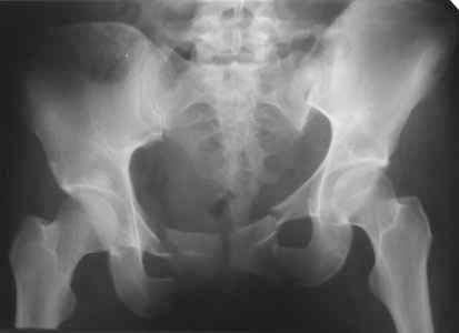
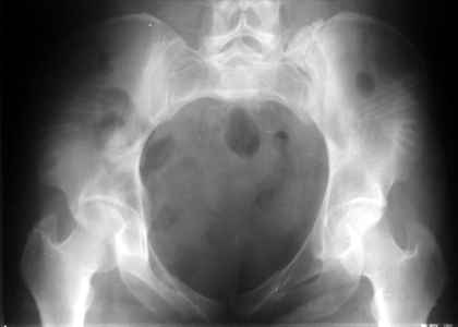
An example of closed ex-fix applcation in a similar case (pelcvic injury with posterior lesion) is attached. I put only AP views to decrease size of the posting.
 |
 |
 |
Best regards,
Alexander N. Chelnokov
Ural Scientific Institute of Traumatology and Orthopaedics
str.Bankovsky, 7. Ekaterinburg 620014 Russia
Date: Fri, 20 Apr 2001 19:15:38 -0500
From: Kyle Dickson, MD
Tom,
I agreed with you up until the end. The most anatomical reduction of the pelvis I believe in this case is from the back because you can use a variety of clamps and check your reduction where anteriorly you have to hold the reduction while someone fixes it for you. This does require a second stage for reduction of the symphysis. There is no clinical support for fixing the minimally displaced rami fractures and I don't think these should be fixed.
kyle
Date: Fri, 20 Apr 2001 19:32:19 -0500
From: Kyle Dickson, MD
Dear alex,
if these xrays are all from the same case this is a very nice reduction. I would like to see the three views of the pelvis pre and post as well as the ct. kellam study showed 27% of unstable pelvis fractures maintained their reduction with an external fixation. Additionally, this case is through the si joint instead of the crescent fracture that was presented. These behave differently.
kyle
Date: Sat, 21 Apr 2001 11:00:47 +0600
From: Alexander Chelnokov
Hello Kyle,
KDM> Dear alex, if these xrays are all from the same case
THX.
KDM>this is a very nice reduction. I would like to see the three KDM> views of the pelvis pre and post as well as the ct.
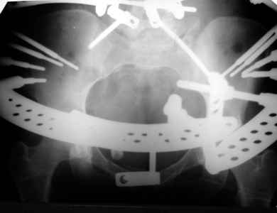
Only inlet views - no outlet and CT were performed in this case.
 |
 |
 |
KDM>kellam study showed 27% of unstable pelvis fractures maintained their reduction with an external fixation.
To perform reduction in these injuries, control on posterior aspects of the pelvis is needed. A circular fixator with half-pins into posterior spinae and/or sacrum is necessary. Was the mentioned ex-fix of this type?
KDM>Additionally, this case is through the si joint instead of the
I just sent a case where i didn't need to prepare images and more or less suitable for the context.
Best regards,
Alexander N. Chelnokov
Ural Scientific Institute of Traumatology and Orthopaedics
str.Bankovsky, 7. Ekaterinburg 620014 Russia
Date: Sat, 21 Apr 2001 9:32 AM
From: Bill Burman, MD
Alex
Thank you for the interesting xray images of the anterior/posterior circular pelvic ex fix.
 |
 |
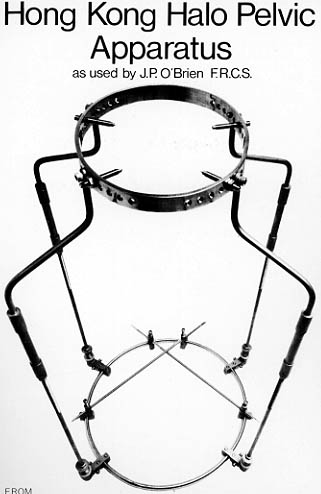
O'Brien and Hodgson used transiliac pins (which could wander into the abdominal cavity) but I always wondered whether half pins posteriorly (as has been applied in your case) could mechanically do the job with less risk.
If they can (and it seems they did in your case of an SI joint disruption which is probably a bigger challenge to obtain and maintain reduction than a crescent fracture), what about
Do you have a clinical photograph of such a pelvic ex-fix on a patient which you could send along?
Bill Burman HWB Foundation
Date: Sat, 21 Apr 2001 10:23:12 -0500
From: Adam Starr
I would plate the symphysis, stabilize the right superior ramus fracture with a screw, and fix the iliac wing fracture with 2 perc screws. I bet the left SI joint is okay.
If good fluoro wasn't available, or if perc techniques made me sick, I'd plate the iliac wing fracture, as described by Dr. J. Borelli. While plating, I'd look for devitalized tissue - common with this injury pattern - and debride it, as described by Dr. Chip Routt.
Then I would get the patient up with crutches, foot flat weight bearing for 3 months.
Adam Starr
Dallas, Texas
Date: Sun, 22 Apr 2001 01:31:53 +0600
From: Alexander Chelnokov
Hello Bill,
BB> O'Brien and Hodgson used transiliac pins (which could wander into the abdominal cavity)
There are methodics with transiliac wires and circular frames. Despite there is no problem to insert the wires without abdominal perforations the frame is not stable enough to control motions in frontal plane (rotation of hemipelvis around AP axis). Though we used it for 3-4 years in late 80-s and early 90-s (see attached scheme).
 |
BB>but I always wondered whether half pins posteriorly (as has been applied in your case) could mechanically do the job
In combination with iliac ones - definitely.
BB>with less risk.
Never met such complications.
BB> what about 1) abdominal access for the general surgeons
Well, the definitive assembly can be applied after stabilization of patients's condition and even later. For acute settings a more simple anterior frame is more suitable. Recent years we rarely meet acute patients, mostly they a referred to us after 1-4 weeks and even more :-( of initial treatment in other hospitals, and laparotomy/cystostomy was done prior to admission to us.
BB>2) posterior pin placement technique and time
When all needed in supine position is done (half-pins to iliac crest and frame assembly) patient is turned on left/right side and half-pins are inserted into posterior aspect. Technique is usual - stub wound, drill and/or awl, and 6 mm titanium half pin.
BB>3) posterior pin tract infection rates
Not outstanding.
BB>4) the need for special beds
Yes, a gap for the device is needed.
BB>and nursing
I am not sure what is meant as nursing but patients can ambulate at least with crutches then with full weigt-bearing so ability of self-care is restored early. We meet them monthly to check x-rays and decide about hardware removal, and they usually change pads around pins in their local facilities.
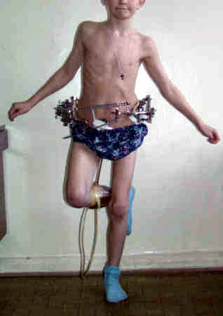
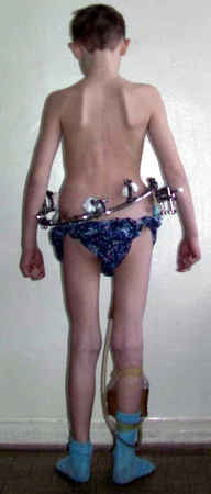
BB>Do you have a clinical photograph of such a pelvic ex-fix on a patient
AFAIR i already sent such a photo to this list or to orthopod, don't remeber when. OK, i'll check for the photo this Monday.
Best regards,
Alexander N. Chelnokov
Ural Scientific Institute of Traumatology and Orthopaedics
str.Bankovsky, 7. Ekaterinburg 620014 Russia
Date: Tue, 24 Apr 2001 03:12:35 +0600
From: Alexander Chelnokov
Hello,
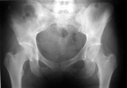
Some images of the subject.
 |
 |
 |
 |
Best regards,
Alexander N. Chelnokov
Ural Scientific Institute of Traumatology and Orthopaedics
str.Bankovsky, 7. Ekaterinburg 620014 Russia