
Date: Tue, 20 Mar 2001 16:10:14 -0000
Subject: Pelvic Fracture
From: Dr. Josep M. Muñoz Vives
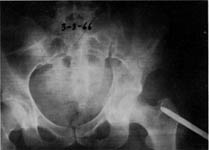
This 19 yo lady substained a fracture of the pelvis and a femoral fracture the day before yesterday attempting suicide. No associated toraco-abdominal lesions. No brain lesion.
|
It is scheduled for surgery next monday. My intention is to fix the acetabular fracture with a plate and the femoral fracture with a nail on the same day.
My doubts are:
What should be done first:
A) The pelvic fracture or the femoral fracture ?
B) Should the sacral fracture be fixed keeping in mind that:
- 1) it is a compression fracture
- 2) there is no vertical displacement
- 3) The reduction maneuvers in the acetabular fracture are capable to displace it???
Thanks in advance
Dr. Josep M. Muñoz Vives
Orthopedic Dept.
Hospital Universitari Dr. Josep Trueta.
Girona
Catalunya, Spain
Date: Tue, 20 Mar 2001 10:38:28 -0800
From: Chip Routt
Thanks-
Her hip remains incongruent. Have you located the femoral head beneath the dome yet? Maybe the displaced transverse won't allow stable reduction using closed techniques until Monday. We'd advocate urgent open reduction if it remains displaced.
Examine her pelvic ring...you'll find it to be quite unstable to examination.
We'd prioritize the acetabular accurate reduction and stable fixation initially (the selected exposure depends on the fracture details that you have from his radiographic scans), then stabilize the sacral (and possibly ramus) fracture percutaneously, then antegrade statically locked femoral medullary nailing.
If her femoral head is not stable and congruent beneath the acetabular dome after closed manipulative reduction, you should not wait.
In reality, we wouldn't wait even if it was stable and congruent...why wait?
Fix her and move on.
Keep waiting, and she gets her wish!
Chip
Date: Tue, 20 Mar 2001 12:39:33 -0600
From: Adam Starr
Hi Dr. Vives.
I don't know if there's necessarily a right and wrong answer to any of your questions. What I would do:
The night she came in, I would've fixed the femur with a retrograde nail. I would've waited 'til later to fix the acetabular fracture. Since the nail isn't done yet, you could do the acetabulum first, then do the nail through the incision used to approach the acetabulum. It can be hard to control proximal femur fractures with manual traction, so I hope you have somebody to help pull traction.
I would go ahead and fix the sacral fracture, but there are others who would say it's stable and doesn't need fixing. It's really kind of hard to define pelvic fracture "stability". It would be nice to see inlte/outlet views to see if it's truly non-displaced. I would fix it with iliosacral screws.
Good luck.
Adam Starr
Dallas, Texas
Date: Tue, 20 Mar 2001 13:55:31 -0500
From: Kevin Pugh
Adam,
I am with you. I can't figure out why the femur isn't fixed yet. I would have fixed the femur with a retrograde nail in the first 24 hours. At our institution, it would have been done by the guy on call, with the pelvis lateraled to the trauma service in the morning.
I would like to see more CT cuts/films etc, but I would put a screw across the sacral fracture. The front could be done with ex-fix or perc screws depending on your ilk.
The acetabulum obviously needs to be fixed. The question is whether to do the pelvis or the acetabulum first. There probably isn't a correct answer, but many preferences I'm sure.
As it is now, and at one sitting, I would do the femur (a stabilized femur may help when fixing your tab), then pelvic ring, then acetabulum.
KP
Date: Tue, 20 Mar 2001 13:27:40 -0600
From: Steven Rabin
Fix the femur by retrograde nail on the day she comes in. Use a retrograde nail because with the medial displacement of the acetabular fracture, it will be difficult to get your awl or guidewire into the pyriformis fossa because you will be hitting the pelvis or the patients body. A retrograde technique allows easy access to the femur. Fixing the femur first allows you to pull traction through the femur during your acetabular reconstrruction. If you do the acetabulum first, it may be more difficult to pull the femur lateral and down without a trochanteric pin. (On the other hand, if you do the acetabulum first, you can do an antegrade femur rod at the same time through the same incision, but overall i think it is easiest to do the femur first even though it requires a separate approach at the knee)... in either case, i'd hate to wait a week to fix them due the risks of prolonged bedrest (pulmonary, decubiti, thrombotic problems etc...).
Date: Tue, 20 Mar 2001 13:44:30 -0600
From: Adam Starr
The reason I'd hate to wait is 'cause the femoral head's grinding on that broken acetabulum. Chip's right - she needs to be fixed quick. You can see the dome of the femoral head on the CT cut. She needs skeletal traction until she gets fixed.
My reason to do a retrograde nail the night of admission would be to avoid making a skin incision around the hip that might compromise my acetabular approach. If I fixed the femur the night of injury, I would put the patient in traction, then fix her acetabulum and pelvis when I had the right team in the OR. At our hospital, the "right team" isn't there at night. You can do acetabulum ORIF's at night (I've done it) but it sucks.
Adam Starr, Dallas, Texas
Date: Tue, 20 Mar 2001 18:16:13 -0800
From: Chip Routt
So, let's try this - pretend you're the patient.
We assume that you understand that the acetabular fracture is the most intolerant injury of the three. If you don't get this, read no further.
If that makes sense, then:
Please then consider the impact of even a minor pelvic ring "fixed" malreduction on your subsequent acetabular reduction attempt.
You also must understand that the dome (cephalad) component of your acetabular fracture is displaced along with and due to the displacement of the ipsilateral sacral fracture. If the sacral fracture is unstable, then this is a "behavioral" associated both column injury. Make sense so far?
Please then reconsider your surgeon's retrograde nailing offer, and its impact on your femoral head (which currently still resides within your displaced acetabular fracture rather than under its dome...this is quite uncomfortable for you and further destroys your joint) and acetabular fracture surfaces.
Do you really want your "first" surgeon to be the "guy on call", who then further destroys your femoral head since it remains displaced (but he/she doesn't have to worry about your acetabulum since he/she won't be responsible for its care), as he/she bangs away "performing" a nocturnal retrograde nailing procedure...you want that for yourself? You don't mind a nail going through your knee? No problem? I don't believe it.
Then, you prefer your displaced sacral fracture to be "lateraled" to your second surgeon for percutaneous fixation, without reduction? It's OK with you if your right hemipelvis is stabilized internally rotated 10-15 degrees?
And then later, you finally get your femoral head gathered up under the dome, but it really doesn't fit too well now since the cephalad component of the acetabular fracture has been malreduced and fixed, and the head is abraded severely from its persistent displacement within the fracture surfaces which are blunted from the same, and the articular acetabular reduction remains "off a bit" due to the above.
So, you look at your final radiographs and see an acetabular malreduction covered in plates, resultant hip incongruity, a sacral malreduction, an abraded head, and a retrograde nail? You vomit.
We're assuming that you've got safe implants.
Your surgeon says, "it was a very bad and complex injury, hopefully it won't get infected."
How are you feeling?
Polytrauma is challenging. Complex inter-related injuries should not be simplified to individual injuries treated independently and routinely, but rather treated considering each injury's impact on the others, and therefore in harmony.
This patient's scenario requires more planning than we can devote based on the shared images. But please, consider how these injuries relate with one another, and then the consequences of your plan(s). Think routinely and creatively.
We all know that not every femoral shaft fracture has to be nailed within 12-24 hours. This is no ordinary patient with a femoral shaft fracture.
I don't want to vomit, so please accurately reduce my acetabular fracture soon using an appropriate exposure, and make it stable to allow early motion. Then reduce and stabilize my pelvic ring injury(ies), before you reduce and stabilize my femur. Combine exposures if you can. Please stay out of my knee unless there's just no other way. Be smart, be quick, and be strategic. Get me up, I want to get back to work.
Respectfully-
Chip
Date: Wed, 21 Mar 2001 15:20:28 -0600
From: Steven Rabin
ok, good point. i was in favor of retrograde rodding but i agree that pounding the rod in, no matter how gentle the surgeon tries to be, is not going to be good for the femoral head. i still think that both need to be done sooner rather than later. If the surgeon is not comfortable with treating the acetabulum at the same time as the femur, then he probably should transfer it to someone who does feel comfortable. If it is impossible to transfer it, and the patient must wait to have the acetabulum done, then i would still do the femur - although it wouldn't get the patient out of traction which would still be needed for the pelvis, fixing the femur might decrease the incidence of fat emboli and make the patient more comfortable. Plating the femur using minimally invasive techniques if possible would avoid the pounding on the femoral head while still stabilizing the femur. Of course it would help to see some femur x-rays.
Date: Wed, 21 Mar 2001 18:04:36 -0500
From: Kevin Pugh
Very nice hyperbole.
First, I would hope that even the 2nd year ortho resident knows that a gentle pull on this leg brings the femoral head under the dome. So, since we are not operating in a vacuum, and I assume that someone is driving the ship, that this gets done in my hospital.
Two, with the head under the dome, and, knowing that just sitting there I have forces of 7x body weight across the joint and it is not in some forceless dream world..and the fact that my femur is moving all over the place and hurting like hell, keeping me in one position, taking a bunch of pain meds, breathing less deeply, and getting atelectasis and whatever else...fix my femur as atraumatically as you can. I know that the guys who take call at our hospital can do this. To really manipulate the hip, the femur should be in continuity.
Three, I don't believe anyone thought that fixing the sacrum in situ was a good idea. I guess there was an implied reduction.
"Polytrauma is challenging. Complex inter-related injuries should not be simplified to individual injuries treated independently and routinely, but rather treated considering each injury's impact on the others, and therefore in harmony."
I agree. It is more than just this acetabulum.
KP
Date: Thur, 22 Mar 2001 15:06
From: Bill Burman
Can intra-operative or even perioperative lateral femoral traction
 |
be protective of the femoral head in the event of a retrograde femoral nailing or a delay in this case?
Bill Burman, MD
HWB Foundation
Date: Fri, 23 Mar 2001 08:02:52 -0500
From: E F Barrick
Bill,
Not in my experience. I was just consulted on a patient in traction and his injured extremity was in a cross-legged position with the foot under the contralateral leg. There is usually wear on the femoral head on those cases that are delayed for some reason. The last one I had to wait 4 weeks and had to do a primary total hip.
E. Frederick Barrick, MD
Director of Orthopaedic Trauma
Inova Fairfax Hospital
Falls Church, VA 22042
Date: Tue, 27 Mar 2001 10:10:00 -0800
From: Chip Routt
Thanks Bill-
It's a good question.
Unlike Dr. Pugh's junior residents' experience (which I assume is quite vast at the R2 level), the rest of us are not always able to achieve congruent and stable reduction of the femoral head beneath the dome, ESPECIALLY in certain transverse acetabular fracture with displacement patterns, as presented...some call it "central fracture dislocation", others say "traumatic protrusio", and on and on.
Toss in the ipsilateral shaft fracture, and we're beyond the "gentle pull" concept.
Again, just stop here if this makes no sense to you.
The caudal/mobile segment of transverse fracture patterns typically depends on symphyseal mobility (and other factors) in order to displace...in this patient, the clinical situation is further complicated by the contralateral comminuted and displaced ramus fractures (an additional "hinge" for the mobile caudal segment) , as well as the displacement of the cephalad segment (which is typically nondisplaced and stable, but for this patient is displaced and has potential rotary instability).
The femoral head follows the caudal segment of the transverse fx as it displaces, likely due to residual capsular tissues and the ligamentum teres remaining intact...this is NOT applicable in those patients with posterior dislocation, or transverse with associated posterior wall fracture patterns - which have lost these soft tissue connections.
Lateral proximal femoral traction is easy and rapid, and can be attempted in this situation...the head may not remain stable unfortunately due to the size of the intact dome, and/or its displacement which also contributes to "coverage". If there's excellent dome coverage...for example an infratectal transverse pattern...then it's possible to achieve and maintain excellent preoperative sphericity of the joint without sustained lateral traction...ie-the head has an adequate "home" or coverage.
In juxta- and trans-tectal transverse patterns, all bets are off since their behaviors are dependant on a variety of factors...think about the transtectal fracture's anterior fracture line as it divides the anterior column at the iliopsoas gutter...now think about the tendon's displacement into the fracture site...ever try to remove it, then reduce and just clamp the fracture - even using an open exposure...was that easy for you?
Sure it was, no problem.
Just keep on working (and thinking while you work) and you'll see what I mean.
We just don't know for this individual patient, but as presented she remains displaced which is not good for her joint's future...think she was depressed before she junmped, stay tuned for her depression with a crummy hip. She's going to perk right up.
We do know that the hip doesn't "take a joke"...and in most situations demands priority, and always perfection.
It's reasonable to attempt urgent closed reduction, with or without sustained lateral traction for this patient. It may or may not work, but it's good to try.
It's unreasonable to "lock in" to simplistic treatment methods for complex patients.
Chip
Date: Wed, 28 Mar 2001 16:48:08 -0000
From: Dr. Josep M. Muñoz Vives
Thank you all for your comments and suggestions.
The lady was operated last Friday (5 days after the fracture). I've waited for the postoperative CT to show you the results.
What I did was:
 |
a) Reduce the acetabular fracture through an ilioinguinal approach. I don't think the head could be reduced by closed means because it was quite hard to reduce the fracture, and harder to maintain it reduced.
By the way what are the list suggestions about holding the reduction in transverse fractures. If you use a clamp in the pelvic brim it annoys when you place the plate. If you use a Matta clamp from the inferior part of the obturator foramen to the anteroinferior iliac spine, which I did, then you are putting a great pressure on the vessels.
No pressure necrosis of the cartilage was seen in the part of the femoral head that could be glimpsed through the fracture, but maybe there was some in a more lateral part, or maybe the condylar traction protected it (don't think so).
b) I fixed the sacral fracture with an iliosacral screw. It's hard to know how stable is a sacral fracture when you don't have an intact femur and no "push-pull" fluoro can be done, and oneself is a donkey and tries to obtain them after closing the ilioinguinal approach and the pelvis can't be held, so I decided that it "could" be unstable and fixed it.
c) I fixed the femoral fracture with an antegrade nail. Although the lady was quite obese and I am a retrograde nail supporter, I agree that I don't know what will happen to retrograde nail knees.
d) Maybe I should have fixed the pubic ramus, but I've no experience with retrograde pubic ramus screws and thought that open fixation had more risks than advantages in this case. Now she's sitting outside of bed and doesn't complain of pain in her left side. Whom of you would let her walk with partial weight bearing on the right side?
A last question: When this lady arrived, although there was no hemodynamic instability, we transfused her 4 units of packed red blood cells. And the day after her fall the Quick was 60%. The day I operated her, the Quick was 80%, that's one of the reasons why I've waited. Do you think that early operation means more blood loss or not?
Dr. Josep M. Muñoz Vives
Orthopedic Dept.
Hospital Universitari Dr. Josep Trueta.
Girona
Catalunya, Spain
Date: Wed, 28 Mar 2001 11:22:23 -0800
From: Chip Routt
a) Reduce the acetabular fracture through an ilioinguinal approach. I don't think the head could be reduced by closed means because it was quite hard to reduce the fracture, and harder to maintain it reduced.
Imagine that...see our previous notes regarding this patient.
By the way what are the list suggestions about holding the reduction in transverse fractures. If you use a clamp in the pelvic brim it annoys when you place the plate. If you use a Matta clamp from the inferior part of the obturator foramen to the anteroinferior isquial spine, which I did, then you are putting a great pressure on the vessels.
A variety of clamp applications (after reduction) are possible which provide stability during fixation without vascular distortion. If you extended your incision medially (I'm assuming that you didn't based on the skin staple line) to include the Stoppa window, you'd be pleased with the improved visualization of the posterior column component of the transverse, as well as the improved clamp opportunities.
Your implant spans the anterior column portion of the transverse, but I see no lag screw across the fracture, nor long screw fixation into the posterior column portion...both techniques improve stability in lab testing.
No pressure necrosis of the cartilage was seen in the part of the femoral head that could be glimpsed through the fracture, but maybe there was some in a more lateral part, or maybe the condylar traction protected it (don't think so).
It's difficult to see the superolateral head thru this fracture line unless you leave it displaced during viewing.
Look at your dome axial postoperative image ... see the acetabular side fracture impaction injury adjacent to the dome medially?...also see the minimal residual fracture distraction? these two situations combined cause the femoral head to be incompletely centered beneath the dome (seen on the 3rd axial postop image)...see how the head is only slightly medially located relative to the dome???
Through this exposure, using a preferential anterior clamp application, the reduction is "eccentric", and this is the result. The transverse fracture's reduction must be "balanced", and then similarly balanced fixation supports such a reduction...the anterior plate as shown does not provide balanced fixation for this particular fracture pattern.
b) I fixed the sacral fracture with an iliosacral screw.
I'm so proud of you for doing so.
This was a 3 column sacral fracture.
The screw has been inserted after an acceptable reduction. If you had chosen a starting point 3mm more posteriorly, even with the same aim, then you would have avoided her SI articular surfaces and maintained the same balanced implant length ... see the sacral postop CT axial image ... is the left sided sacral linear lucency at the screw tip an artifact, or fracture that we initially didn't identify?
c) I fixed the femoral fracture with an antegrade nail.
God Bless You. You are a kind and thoughtful surgeon and physician.
Maybe an "only slightly more lateral" starting point avoids potential problems.
Is her rotation symmetrical?
d) Maybe I should have fixed the pubic ramus ... Whom of you would let her walk with partial weight bearing on the right side?
Depends on symptoms...if she has no pain with full left sided WB, then sure I'd advocate protected "weight of limb" WB for her right side.
A last question:
Do you think that early operation means more blood loss or not?
We've found no difference statistically, using before or after postinjury day #3 as the factor.
Thank you very much for identifying an interesting and educational patient for discussion.
Thank you for sharing the studies both pre- and postoperatively.
Please review the postop plain films and be attentive to the femoral head's relationship with the dome...even minimal residual displacement of the unstable caudal segment yields head-dome congruity problems...do you see it?
Share her outcome later-
Chip